

This guide explains endocarditis symptoms, the diagnostic pathway, treatment decisions and prevention. It focuses on infective endocarditis and includes practical questions to discuss with the clinical team.
What endocarditis means
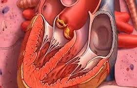
Endocarditis affects the inner surface of the heart, especially its valves. In infective endocarditis, microorganisms establish an infection there, sometimes forming attached material called a vegetation. This material can damage a valve or release fragments into the circulation. Most infections are bacterial; other organisms can occasionally be responsible. A native valve is a person’s own valve, while a prosthetic valve is a replacement. Infection can also involve implanted cardiac material. These distinctions matter because the tests, complications and treatment plan can differ. This guide concentrates on infective endocarditis, a serious illness that needs professional assessment and coordinated treatment. [8]
Not every valve vegetation represents an infection. Nonbacterial thrombotic endocarditis involves deposits associated with an underlying condition, such as cancer or an autoimmune disorder. It can produce emboli, including strokes, while blood cultures remain negative. A scan showing a mass therefore does not, by itself, identify its cause. Equally, a negative culture does not establish that a mass is noninfectious. Clinicians interpret the person’s symptoms, laboratory results, imaging and medical background together. Distinguishing these possibilities is essential because treatment aimed at an infection and treatment for a noninfectious clotting process are different. Neither diagnosis should be made from an online symptom checklist. [17] [5]
Risk depends on the heart and the route into the bloodstream
People with a previous episode of endocarditis, certain congenital heart conditions, damaged valves or artificial valves have particular reasons to discuss risk with their clinical team. Some cardiac devices also change the assessment. Risk is not a yes-or-no label: the kind of heart abnormality, the presence of implanted material and other health problems all matter. Endocarditis can also occur in someone without a previously recognized heart condition. Having a risk factor does not mean every fever is endocarditis, but it is useful information when reporting an unexplained illness. Keep the exact name of a valve procedure or implanted device in your health records. [44]
The organisms involved are not identical in every setting. Studies of patients with definite endocarditis have identified staphylococci, streptococci and enterococci among important causes, with patterns influenced by healthcare exposure and the population being studied. A recent hospital stay, an infected skin wound, a vascular catheter or another bloodstream infection can help shape the investigation. The clinical team is trying to find both the organism and the place from which it entered the bloodstream. That history guides testing; it cannot reliably identify the cause on its own. Avoid assuming that a dental procedure explains an infection simply because it happened before symptoms began. [25] [44]
Symptoms can begin gradually or deteriorate quickly
Fever, chills, sweats, fatigue and feeling generally unwell can develop over days or weeks. Some people notice poor appetite, weight loss, muscle or joint discomfort, or breathlessness. These symptoms overlap with many other illnesses, so the pattern and medical background matter more than any single symptom. Small skin or nail changes described in textbooks are neither required for diagnosis nor a dependable home test. Seek prompt assessment for a persistent, unexplained fever, particularly with a previous episode, a replacement valve or other recognized cardiac risk. Tell the clinician when symptoms began and whether antibiotics have already been taken for this illness. [9]
Sepsis is a life-threatening response to an infection and can occur with endocarditis. Warning signs include new confusion, difficulty breathing, clammy or sweaty skin, severe discomfort, a fast heart rate or a weak pulse. Someone may feel exceptionally cold or have fever and shaking. Do not wait for all these features, for a particular thermometer reading, or for a routine appointment when the person is deteriorating. Use the local emergency service and explain that a serious infection is possible. Emergency assessment should take priority over arranging a dental review, gathering old records or trying to obtain particular tests independently. [10]
A fragment released from an infected valve may interrupt blood flow to the brain. Sudden face drooping, weakness or numbness on one side, trouble speaking or understanding, new vision problems, loss of balance, or a sudden severe headache require emergency help. Note when the person was last known to be well and tell the emergency team about suspected or confirmed endocarditis. Symptoms that disappear still need urgent assessment because a temporary improvement does not settle the cause. Do not drive yourself with possible stroke symptoms. The stroke team needs to assess treatment options in the context of the infection and any bleeding risk. [11] [41]
Valve damage can affect how effectively the heart moves blood forward. Breathlessness at rest, difficulty breathing when lying flat, waking suddenly short of breath, or increasing swelling should be reported promptly. Severe breathing difficulty, collapse or a rapid deterioration needs emergency care. A person may mistakenly attribute reduced stamina to being tired from the infection, so a clear change from their usual breathing deserves attention. The team may assess circulation, oxygen levels and valve function rather than treating the symptom in isolation. Do not change a prescribed heart medicine or fluid plan without advice from the clinicians responsible for your care. [20] [26]
Diagnosis combines blood tests, imaging and clinical judgment
Blood cultures and a useful history
Blood cultures are samples taken to look for microorganisms in the bloodstream. Clinicians usually aim to obtain appropriate cultures before antibiotics when this can be done promptly and safely, because treatment can reduce the chance of identifying the organism. The timing is a clinical decision: urgent treatment must not be delayed in an unstable patient. Do not withhold prescribed antibiotics, stop an existing course or postpone emergency care to improve a test result. Tell the team what medicines were taken and when. Several samples and follow-up cultures may be needed to distinguish persistent infection, a meaningful organism and contamination introduced during sampling. [35] [48] [21] [1]
There is no single test that settles every case. The Duke-ISCVID diagnostic criteria bring together microbiology, heart imaging and other clinical findings, and their 2023 revision recognizes newer molecular and imaging methods. These criteria help clinicians classify the evidence; they do not turn an uncertain early presentation into a simple home score. Information can change as cultures develop or additional images become available. A person may initially have a possible diagnosis while the team continues investigating. Ask which findings support infection, which are still uncertain and what the next test is meant to resolve. A working diagnosis should be explained in ordinary language. [12]
What ultrasound can and cannot show
An echocardiogram uses ultrasound to examine the heart. A transthoracic examination looks through the chest wall and is often an early test. A transesophageal examination places the ultrasound probe in the esophagus, closer to the heart, to obtain different views. The latter requires preparation and assessment of procedural suitability. A negative transthoracic study cannot reliably exclude endocarditis in every patient: small lesions, image quality and the structure being examined can limit what is visible. The decision to obtain further or repeat imaging depends on the whole clinical picture. Ask what the scan showed about valve function as well as whether a vegetation was seen. [18] [36] [3]
Replacement valves and other implanted material can make images more difficult to interpret. Artificial components may obscure nearby structures, and an infection may sit around a valve rather than appear as an obvious mobile growth. This helps explain why a reassuring initial ultrasound does not always finish an investigation when suspicion remains high. Patients with a transcatheter aortic valve replacement have additional imaging challenges. The team may compare previous studies, obtain a different imaging method or repeat an examination as the clinical picture develops. Bringing the device or valve information to appointments can help clinicians select the appropriate investigation and interpret what they see. [37]
Cardiac computed tomography can add a detailed view of the tissues around a valve. It can be particularly helpful when clinicians are looking for an abscess or another complication extending beyond the valve itself. CT and ultrasound answer overlapping but different questions; one does not automatically replace the other. A study’s ability to detect a small vegetation is not the same as its ability to map surrounding damage before surgery. Contrast exposure, kidney function and the reason for ordering the scan are considered individually. Ask what additional question CT is expected to answer and how that result could change the treatment plan. [27] [47]
Some patients need imaging that looks for abnormal metabolic activity, such as fluorodeoxyglucose PET/CT. This can support investigation of selected prosthetic-valve or device infections and sometimes reveal sites of infection elsewhere. Preparation and interpretation are specialized. Inflammatory changes related to surgical materials can complicate interpretation, while low inflammatory activity may reduce detection. A positive scan is therefore evidence to interpret alongside cultures and other images, rather than proof in isolation. A negative study also has limits. Its usefulness depends on the clinical question, the valve or device involved, and the expertise available to read the examination. [38] [24] [1]
When blood cultures stay negative
Blood culture-negative endocarditis remains possible when routine cultures do not grow an organism. Prior antibiotics are one explanation; organisms that are difficult to grow are another. The American Heart Association’s 2025 statement emphasizes a structured investigation informed by the clinical history. Travel, animal contact, previous procedures and other exposures can guide specialized blood tests or molecular testing. The team may also reconsider noninfectious explanations. Negative cultures do not justify choosing an antibiotic at random, nor do they prove that treatment is unnecessary. Tell clinicians about earlier antibiotics and relevant exposures without assuming which one caused the illness. [5]
Molecular tests can look for microbial genetic material in blood or tissue, including tissue collected during an operation. They may provide useful information when ordinary cultures are unrevealing, but the type of sample and the test’s performance matter. A result must be interpreted with the clinical and microbiology teams: detection of genetic material is not always equivalent to a living organism causing ongoing disease. These methods complement conventional testing rather than eliminating the need for cultures, careful sampling and pathology. Ask whether a result changes the suspected organism, the confidence in the diagnosis or the treatment plan, rather than viewing every positive result as a separate infection. [31] [42]
Why clinicians look beyond the valve
Vegetations can shed infected material into the circulation. In left-sided endocarditis, emboli may affect organs such as the brain, spleen or kidneys; right-sided disease can send material toward the lungs. The location of pain or other symptoms may therefore lead to tests away from the heart. The risk is influenced by more than the mere presence of a vegetation, and clinicians consider its features together with the organism, previous embolic events and response to treatment. Report new neurological symptoms, sudden severe pain or a new breathing problem promptly. A scan showing a vegetation is not a reliable basis for predicting an individual’s outcome at home. [41] [1]
Heart failure is an important complication because an infected valve may become badly damaged and leak. This can create a mechanical problem even while antimicrobial treatment is controlling the organism. Large clinical cohorts have linked heart failure in endocarditis with worse outcomes, which is one reason it changes the urgency of specialist assessment. Whether an operation is appropriate depends on the severity of the valve problem, the infection and the person’s overall situation. The practical question is not simply whether a culture has become negative, but whether circulation and heart function are improving. Medical and surgical teams consider these issues together. [26]
Endocarditis can sometimes trigger inflammation in the kidney’s filtering structures, a problem called glomerulonephritis. A biopsy-based series documented this complication, but those carefully selected patients do not show how common it is in everyone with endocarditis. Changes in kidney function or urine findings need clinical interpretation; they should not be assumed to have the same cause in every patient. The team may need kidney expertise to understand the problem alongside the infection. Ask what the results mean and how they affect monitoring. Do not start treatment for inflammation or alter the infection plan without specialist advice. [33]
Treatment is a coordinated clinical plan
The American Heart Association’s 2026 statement emphasizes an Endocarditis Team. Depending on the case, this can bring together cardiology, infectious diseases, cardiac surgery, microbiology, imaging specialists and other clinicians. Their decisions are linked: an imaging finding may change the need for surgery, and identifying the organism may change antimicrobial treatment. The team should also explain who coordinates care and whom to contact between reviews. Shared planning is particularly valuable when tests disagree or when the safest timing of a procedure is uncertain. A clear explanation of the current diagnosis, immediate priorities and next decision helps patients participate without having to reconcile separate specialist opinions themselves. [2]
Antimicrobial treatment is selected for the organism, its susceptibility, the valve or device involved and the person’s clinical circumstances. Initial treatment may begin before the cause is fully identified, then change when reliable results return. Many patients receive intravenous therapy, and treatment commonly extends over several weeks. The precise medicines, route and duration are individual clinical decisions. An online drug list cannot account for kidney function, allergies, interactions or surgical findings. Take the prescribed plan seriously even when fever improves. Report a missed dose, vomiting that prevents a dose being taken, a new rash or another concerning effect promptly so the team can advise safely. [46] [21]
Treatment requires monitoring as well as giving the medicine. The team may repeat blood counts, kidney or liver tests and cultures, depending on the treatment and clinical course. Intravenous access also needs care because a catheter can develop its own complications. New pain, redness, swelling, leakage or difficulty using the line should be reported through the service’s instructions. Know which symptoms require immediate help and which contact can give same-day advice. A written schedule is useful when several services are involved. Laboratory results, line checks and review appointments are part of treatment, even when they happen outside the hospital. [15] [23]
When surgery or device removal enters the discussion
An operation may be considered for severe valve dysfunction with heart failure, infection that cannot be controlled adequately, or substantial concern about further emboli. These reasons can overlap. The purpose may include removing infected tissue, repairing damage and restoring effective valve function. Some situations need urgent surgery before the antimicrobial course is finished; other cases can be managed without an operation. The decision weighs expected benefit, procedural risk, complications and patient preferences. Ask which problem an operation is intended to solve, how urgent that problem is and what may happen if the procedure is deferred. There is no single timetable that fits every patient. [3]
Research on early surgery helps inform these discussions but has limits. A randomized trial in selected patients with left-sided endocarditis, severe valve disease and large vegetations found benefit for an early-surgery strategy on its combined outcome, driven largely by fewer embolic events. That result cannot be applied automatically to every person with a vegetation or to every type of endocarditis. Trial participants met specific selection criteria and received specialist care. For an individual, the team must still consider heart failure, the organism, brain complications and operative risk. Ask how closely the evidence being discussed matches your own clinical situation. [14]
A stroke or bleeding in the brain can complicate the timing of valve surgery. Delaying an operation may allow the heart or infection to worsen, while operating can carry neurological risks. The answer depends on what happened in the brain, the severity of the neurological injury and the urgency of the cardiac problem. Studies of surgery after neurological complications help teams assess those tradeoffs, but they do not supply a universal waiting period. Brain imaging and joint discussion between cardiac, surgical and neurological specialists are important. Patients and families should ask which risks are most immediate and what findings would change the recommended timing. [39]
An infected pacemaker or defibrillator system raises different questions from an infection limited to a native valve. The generator pocket, leads and bloodstream may all need assessment. For definite device infection, specialist teams generally recommend complete system removal alongside antimicrobial treatment. Exceptions require individual assessment when removal is unsuitable. Extraction is a specialist procedure; patients should never try to manipulate a device or manage an inflamed pocket themselves. The team also plans whether another device is needed and how that need will be met safely. Device-related fever, pocket changes or new illness should be reported promptly, even years after implantation. [24]
Treatment outside hospital is for selected patients
The POET trial studied a switch to oral antibiotics in carefully selected, clinically stable adults with certain forms of left-sided endocarditis after an initial intravenous phase. Its findings support an option within a structured specialist pathway; they do not mean that tablets can replace initial hospital care or suit every organism, valve or complication. Before a switch, the team must be confident that the person and treatment plan meet appropriate criteria. Oral treatment still requires reliable access to medicines, review and monitoring. Do not convert a prescription to tablets, use leftover antibiotics or shorten treatment because the symptoms have improved. [13]
Treatment duration is also being studied. The 2026 POET-II trial compared a clinician-tailored shorter course with standard duration in selected, stable patients with left-sided endocarditis after initial treatment. The tailored strategy met the trial’s statistical criterion for its combined safety outcome, but relapse was more frequent. These findings need careful interpretation and do not give patients permission to stop antibiotics early. A team considering this evidence must assess how closely a patient matches the study and what follow-up is available. The duration of treatment remains an individual, prescribed clinical decision. [34]
Outpatient intravenous treatment also requires selection and organization. Clinical stability is only one part of the decision: the service must be able to deliver treatment, monitor results, care for the line and respond quickly if problems arise. Studies of outpatient pathways describe outcomes in selected patients receiving this support, rather than showing that all patients can safely leave hospital early. Before discharge, confirm who gives each treatment, where blood tests happen and which number works outside normal hours. Transport, housing, caregiving and cost barriers belong in the plan. Tell the team about practical obstacles early so they can help arrange a workable approach. [43]
Care should include support for substance use when relevant
For a person who injects drugs, endocarditis care should address the infection and substance use together, with respectful, nonjudgmental treatment. The American Heart Association’s statement on this group emphasizes multidisciplinary care that includes addiction expertise. Decisions about surgery and antimicrobial treatment require individual assessment rather than assumptions about someone’s future behavior. Discuss pain, withdrawal, treatment preferences and barriers to staying in care openly with the team. Evidence-based treatment for opioid use disorder and harm-reduction support can be offered alongside cardiac care. People deserve clear explanations and a plan that recognizes their circumstances, including unstable housing or difficulty reaching appointments. [6]
Observational research has examined how addiction consultation and treatment relate to completing antimicrobial treatment and returning to hospital after serious injection-associated infections. These studies cannot prove that one service alone determines an outcome, but they support treating access to addiction care as a meaningful clinical issue. A referral should lead to an achievable next step, such as an identified clinician or service and a follow-up arrangement. If a plan is difficult to follow, explain what is getting in the way before leaving care. A collaborative response can address withdrawal, medication access or other needs that might otherwise interrupt treatment. [28]
Recovery and follow-up continue after the acute infection
A later episode of endocarditis may represent relapse of the original infection or a new infection. Studies distinguish these possibilities using the organism, timing and clinical findings; symptoms alone cannot tell them apart. A previous episode is therefore important information whenever new unexplained fever or other concerning symptoms develop. Contact the treating service promptly rather than starting leftover antibiotics, because fresh assessment and appropriately timed testing may be necessary. Keep a record of the organism, affected valve or device, treatment dates and any operation. These details make future assessment more efficient and help clinicians compare a new illness with the earlier episode. [30]
Recovery is not measured only by the last antimicrobial dose. Research following survivors has described ongoing effects on physical function, fatigue and quality of life. Some people need time and support to return to daily activities or employment, particularly after surgery or a long hospital stay. Discuss persistent exhaustion, sleep problems, anxiety or difficulty managing everyday tasks at follow-up rather than assuming they must be endured. The team can assess whether symptoms reflect recovery, a cardiac problem or another issue. Ask for advice about activity, work and rehabilitation that fits your treatment and current heart function; someone else’s timetable may not suit you. [29]
Before discharge, clarify the follow-up plan for the heart as well as the infection. You may need clinical review, further imaging or tests, and discussion of longer-term valve or device care. Know which team will arrange these steps and how you will receive the results. If surgery introduced new medicines or changed an existing treatment, ask for an updated list and a clear explanation of monitoring. Tell future clinicians and dentists about the episode. A discharge summary is particularly useful when care is shared between hospitals. Seek advice for recurrent fever or new symptoms rather than waiting silently for the next scheduled appointment. [45]
Prevention focuses on everyday care and specific situations
Regular dental care and attention to oral health are part of prevention. Research has shown that bacteria can enter the bloodstream during everyday activities such as toothbrushing as well as during dental procedures. This does not mean brushing should be avoided. It helps explain why prevention cannot rely only on an antibiotic given before an occasional appointment. Tell your dentist about previous endocarditis, valve surgery and relevant congenital heart disease, and ask how to maintain a practical routine. Seek assessment for dental infection or persistent mouth problems. Dental and cardiac teams can coordinate when the heart history makes the choice of a procedure or preventive treatment more complex. [40] [22]
Under American Heart Association guidance, dental antibiotic prophylaxis is reserved for selected people at highest risk of a poor outcome. Categories include prosthetic cardiac valves, including transcatheter valves and homografts; prosthetic material used for valve repair; previous infective endocarditis; unrepaired cyanotic congenital heart disease, or repaired congenital heart disease with a residual shunt or valve leak at or next to a prosthetic patch or device; and a heart transplant with regurgitation caused by a structurally abnormal valve. Relevant dental procedures manipulate gum tissue or the region around a tooth root, or perforate oral mucosa. The clinician and dentist must verify eligibility and the procedure. A pacemaker or defibrillator alone does not qualify. [4]
Evidence about dental prophylaxis includes large observational studies examining invasive dental procedures and subsequent endocarditis in people at different cardiac risk levels. Such work can identify associations and support targeted prevention, but it does not make every dental visit an indication for antibiotics. The expected benefit must be considered alongside adverse reactions and unnecessary antimicrobial exposure. If prophylaxis is recommended, the prescribing team selects a suitable medicine after checking allergies and other relevant factors. This article deliberately does not give a regimen. Keep an up-to-date account of your cardiac diagnosis and prior reactions so the dentist can make the decision using the correct information. [32]
Recommendations also depend on jurisdiction. The European Society of Cardiology’s 2023 guideline recommends prophylaxis for defined high-risk patients undergoing at-risk dental procedures and addresses selected nondental situations differently from American guidance. A recommendation for one category or procedure should not be generalized to every invasive test. Patients travelling or receiving care in another country should provide their cardiac records and ask the local team which guidance applies. The key questions are whether the heart condition meets the relevant risk definition, whether the procedure is covered and whether other circumstances change the decision. Do not take an antibiotic simply because a procedure sounds invasive. [1]
NICE guidance in the United Kingdom says antibiotic prophylaxis is not routinely recommended for dental procedures; this wording is not the same as saying it can never be appropriate. The Scottish Dental Clinical Effectiveness Programme’s second edition, published in March 2026, provides implementation advice. It recommends or asks teams to consider prophylaxis for specified high-risk cardiac conditions, does not recommend it for moderate-risk conditions, and emphasizes a shared decision for at-risk dental procedures. These differences are a reason to have a documented discussion with the dentist and cardiac team. A prior prescription from another country or an earlier procedure should not be treated as a permanent instruction. [7]
Skin care matters because organisms such as Staphylococcus aureus can enter the bloodstream and sometimes infect a valve. Carrying this bacterium on the skin or in the nose is not the same as having endocarditis. Follow wound-care advice and seek help for a worsening skin infection. If you have a vascular line, report changes around the entry site and follow the service’s care instructions. People who inject drugs can benefit from sterile-equipment services and links to addiction treatment. These measures do not remove every risk; an unexplained new illness still needs assessment in the context of your cardiac history. [19] [9] [23] [16]
Frequently asked questions
What is infective endocarditis?
It is an infection inside the heart, often involving its valves. [8]
Can negative blood cultures rule out endocarditis?
No. Prior antibiotics or difficult-to-grow organisms can leave cultures negative. [5]
Does everyone with endocarditis need surgery?
No. Decisions depend on heart damage, infection control and the risk of emboli. [3]
Can endocarditis be treated with tablets?
Only selected, stable patients can switch under a specialist treatment and monitoring plan. [13]
Does every heart condition need dental prophylaxis?
No. Eligibility depends on the cardiac condition, the dental procedure and local guidance. [4]
Can endocarditis come back?
Yes. New unexplained fever after treatment needs prompt clinical assessment. [9]
Sources
- ESC endocarditis guidance
- AHA 2026 management statement
- ACC/AHA valve guideline
- AHA dental prevention card
- AHA culture-negative statement
- AHA injection-associated care
- NICE and SDCEP prevention advice
- NHLBI endocarditis overview
- NHS symptoms and recovery
- CDC sepsis warning signs
- CDC stroke warning signs
- Duke-ISCVID diagnostic criteria
- POET oral-treatment trial
- Early-surgery randomized trial
- IDSA outpatient IV guidance
- CDC harm-reduction services
- Noninfectious-valve cohort
- MedlinePlus heart ultrasound
- CDC staphylococcal infections
- AHA heart-failure symptoms
- CDC antibiotic safety
- NIH daily dental care
- MedlinePlus catheter care
- AHA cardiac-device infection guidance
- International endocarditis cohort
- EURO-ENDO heart-failure outcomes
- Cardiac CT diagnostic study
- Addiction-consultation outcome study
- Recovery-experience interview study
- Relapse and reinfection cohort
- Blood microbial-DNA study
- Dental-prophylaxis observational study
- Kidney-inflammation biopsy series
- POET-II duration trial
- CDC blood-culture collection
- TTE and TEE comparison
- Transcatheter-valve imaging study
- PET interpretation study
- Stroke and surgery cohort
- Toothbrushing and bacteremia trial
- Embolism-risk prospective study
- Valve DNA after treatment
- Glasgow outpatient-treatment cohort
- Guy’s endocarditis patient guide
- British endocarditis-service consensus
- Coventry endocarditis patient guide
- Radiology contrast safety
- Surviving Sepsis clinical guidance