
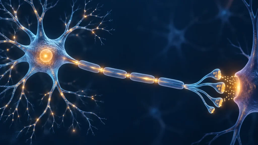
A neuron can receive thousands of inputs without firing, then generate a brief electrical signal when the combined effect is sufficient. That change is often called neuron activation. In a classroom, the phrase usually refers to the path from incoming signals to an action potential and then to communication at a synapse. In a laboratory, “activation” can also describe a change in firing rate, chemical signaling, or activity across a network. The meaning depends on what was measured. It is more precise to ask which part of the process is active than to imagine a neuron as simply switched on or off.
This guide follows a signal from dendrites to axon terminals, explains the electrical and chemical steps without treating one textbook voltage as universal, and connects those steps to behavior and learning. It is written for students who need a reliable conceptual model, particularly those studying biological bases of behavior in AP Psychology. The College Board course framework places the neuron and neural firing in that unit, while the more detailed ion-channel explanation below helps curious readers understand why the simplified terms work. The article is educational, not a way to diagnose symptoms or interpret a person’s brain activity.
Start with the parts of a neuron
Most textbook diagrams show a cell body, branching dendrites, an axon, and terminals. Dendrites and the cell body receive and integrate much of the information from other cells. The axon carries an action potential away from the region where it begins, often the axon initial segment. Terminals can release chemical messengers onto another neuron, muscle cell, or gland cell. This direction is a useful basic map, but real neurons vary considerably in shape and can receive inputs on several compartments. Do not assume every neuron is a perfect drawing with one input end and one output end.
The cell membrane separates the fluid inside from the fluid outside. Charged particles, or ions, do not cross it freely in the same way as water through an open pipe. Membrane proteins provide selective routes, and the concentrations of key ions differ across the membrane. These differences create an electrical potential: the inside is typically negative relative to the outside at rest. The exact value varies among cell types and conditions, so a commonly taught resting value such as about −70 millivolts is an example rather than a number to assign to every neuron.
Glial cells also matter. They support neural tissue, influence the chemical environment, and, in the case of oligodendrocytes and Schwann cells, form myelin around many axons. A neuron does not operate as an isolated electrical wire. Its surroundings affect how it maintains gradients, conducts signals, and participates in a circuit. Sly Academy’s overview of the nervous system provides the larger map of central and peripheral structures in which individual cells work.
Resting potential: ready is not inactive
At rest, a neuron maintains an unequal distribution of ions, especially sodium and potassium, across its membrane. Selective permeability and electrical forces help establish the resting membrane potential. The sodium–potassium pump contributes to maintaining the ion gradients over time by using energy to move ions against their concentration gradients. It is not the sudden switch that creates the rising phase of each action potential. Treating the pump as if it directly fires the neuron confuses long-term maintenance with the rapid event itself.
Imagine a rechargeable system whose stored difference makes a quick response possible. The analogy is limited: the neuron is a living cell with multiple channel types, ongoing metabolism, and constantly changing input. Still, it helps explain why “resting” does not mean “doing nothing.” Channels can be open, local signals can arrive, and gradients are being maintained even when no action potential is traveling along the axon. OpenStax’s action-potential chapter explains the relationship among permeability, ion gradients, and membrane voltage in more detail.
For AP Psychology, learn the idea of resting potential as the membrane’s ready state. The current College Board course description lists resting potential, threshold, depolarization, all-or-none firing, and refractory period among neural-transmission concepts. Its course framework explicitly says that detailed sodium–potassium-pump knowledge is outside the AP Psychology exam scope. The pump is included here as an optional physiological explanation, not as a cue to memorize extra molecular details for that exam. Check your instructor’s expectations if your course covers more biology.
Incoming signals are graded before firing is all-or-none
Communication usually begins with a change at a receptor or synapse. A neurotransmitter binding to receptors may alter ion flow in a small area of membrane. A sensory receptor can be affected by light, pressure, chemicals, or another stimulus, depending on the cell. These local voltage changes are graded potentials: their size can vary with the input, and they tend to weaken as they spread. Some move the membrane voltage toward the level at which an action potential is likely; others move it away or reduce the effect of an excitatory input.
It is helpful to distinguish a graded input from the action potential it may help trigger. “All-or-none” applies to the regenerative action potential once the relevant membrane reaches threshold; it does not mean every synaptic input is the same size or that every tiny stimulus makes the neuron fire. If one weak input alone is insufficient, two inputs arriving close together in time or at different places may combine. Other inputs may counteract them. The neuron integrates an ongoing mixture, not a single vote from one synapse.
Consider a simplified example with no claim that it captures the exact arithmetic of a real cell. A neuron receives an excitatory signal that nudges its trigger region toward threshold, but not enough to fire. A second excitatory signal arrives soon afterward, and the combined effect reaches the level needed to open a self-reinforcing set of voltage-gated channels. If an inhibitory input arrives at the same time, the outcome may change. The lesson is about timing, location, and integration. Real dendrites and synapses are more complex than adding three fixed numbers.
Threshold and the axon initial segment
Many neurons initiate action potentials at or near the axon initial segment, a region rich in voltage-gated sodium channels. When depolarizing input brings this membrane to a critical range, opening of some channels allows sodium ions to enter. That entry depolarizes the region further and promotes opening of more channels. This positive-feedback process produces the rapidly rising phase of an action potential. Below threshold, a local voltage change may fade without producing the full traveling spike.
Textbooks often illustrate threshold near −55 millivolts and resting potential near −70 millivolts. Those values describe a useful example, not an immutable rule for every neuron or every moment. Threshold depends on the membrane’s recent activity and its ion-channel properties; different cells can have different values. When interpreting a graph, focus first on the relationship: resting level, movement toward threshold, the rapid spike after threshold, and recovery. Memorizing a single number without understanding that sequence can lead to wrong conclusions.
Threshold is not a psychological decision made by one cell. It is a property of membrane dynamics. A stimulus can influence a neuron indirectly through other neurons and multiple synapses, and a behavioral response usually requires activity across a circuit. The trigger region is important because it converts integrated graded input into a signal able to travel over a long axon. It does not determine, by itself, the meaning of the stimulus or the person’s conscious experience.
The action potential step by step
Once an action potential starts, voltage-gated sodium channels open quickly and sodium enters. The membrane potential becomes less negative and briefly may become positive relative to the outside. Sodium channels then inactivate, while voltage-gated potassium channels contribute to moving the membrane back toward a negative potential as potassium leaves the cell. Because potassium conductance can remain elevated briefly, the voltage may dip below its usual resting level before returning toward it. This sequence is commonly labeled depolarization, repolarization, and after-hyperpolarization.
Do not picture the same sodium ion racing the entire length of an axon like a ball through a tube. The signal is a wave of local membrane changes: current from one active segment affects the next segment, where channels regenerate the event. Ion movement across the membrane occurs at many successive locations. That is why an action potential can propagate without fading away like a small graded potential. The cell later restores and maintains the gradients that make repeated signaling possible.
The height of a typical action potential in one axon does not grow in proportion to stimulus strength. This is the all-or-none principle. A stronger sustained input can instead change whether a cell fires, how soon it fires, and how frequently it fires within physiological limits. Across a population, it may recruit more neurons. This distinction matters for sensation: a stronger stimulus need not make an individual spike taller; it can alter firing patterns and the number of participating cells. Avoid claiming every spike everywhere is literally identical, because cellular conditions and recording locations can vary.
Why the signal normally travels forward
Immediately after a segment of membrane fires, its sodium channels are inactivated and cannot be opened again in the usual way. This is part of the absolute refractory period, when another action potential cannot start at that segment. During a later relative refractory period, firing is possible but generally requires a stronger depolarizing input because the membrane has not fully returned to its prior state. These phases limit maximum firing rate and help a traveling signal move away from the site just activated rather than bouncing backward.
Refractory periods are not a period in which the entire brain rests. They are brief properties of membrane regions after a spike. Other neurons can still be active, and a single neuron can fire again after sufficient recovery. If a student says a neuron “needs a rest,” that shorthand is acceptable only if it is unpacked into channel recovery and temporary changes in excitability. The purpose is to understand a causal process, not to anthropomorphize the cell.
In a diagram, locate the fresh membrane ahead of the traveling spike and the recently active membrane behind it. The fresh region can respond to current from the active region; the region behind it is refractory. This explains the ordinary forward direction along an axon. It does not mean electrical current itself can never spread backward locally, and it does not forbid carefully designed laboratory stimulation from initiating signals in unusual directions. The basic classroom account describes ordinary physiological propagation.
Myelin changes conduction, not the size of the message
Many axons are wrapped in myelin, a layer made by glial cells. Myelin reduces current loss across covered portions of the membrane. Gaps called nodes of Ranvier contain many channels where the action potential is regenerated. In saltatory conduction, local current spreads beneath the myelin between nodes and the signal is renewed at successive nodes. The everyday phrase “the impulse jumps” is a teaching shorthand; the signal does not teleport across an empty gap.
Myelination and axon diameter influence conduction speed, but faster is not the same as stronger or more meaningful. Some axons are unmyelinated and still communicate effectively for their tasks. The nervous system contains pathways with different timing needs. In a reflex, swift signaling can help coordinate a rapid response; in other contexts, integration across many neurons matters more than maximal speed. Do not judge a brain function from myelin alone.
Damage to myelin can disrupt signaling, but symptoms and causes require clinical evaluation. The AP Psychology framework uses disorders such as multiple sclerosis as examples of how disrupted neural transmission can affect functioning. A student should connect the concept to impaired conduction without treating an educational diagram as a diagnostic test. The page explains mechanisms; it does not imply that fatigue, tingling, or any other symptom proves a particular neurological condition.
Reaching a synapse: electrical to chemical to electrical
When an action potential reaches an axon terminal at a typical chemical synapse, it depolarizes the terminal membrane. Voltage-gated calcium channels open, calcium enters, and vesicles release neurotransmitter into the small synaptic cleft. Molecules diffuse across and bind receptors on a target cell. Receptor activation can change the target cell’s membrane properties, creating a graded postsynaptic response. This is why a signal can be electrical along the axon, chemical across a synapse, and electrical again in a receiving cell.
Not every synapse is chemical, and not every neurotransmitter has one simple “excitatory” or “inhibitory” identity in all settings. The outcome depends on the receptor, cell, and circuit. A chemical synapse has steps that take time and allow modulation. An electrical synapse connects cells through gap junctions and can pass current more directly. Most introductory psychology discussions emphasize chemical synapses because neurotransmitter release, receptors, and clearance help explain many familiar learning and drug concepts.
After release, neurotransmitter does not remain in the cleft forever. It can be taken back up by cells, broken down by enzymes, or diffuse away, depending on the messenger and synapse. Reuptake is one possible clearance mechanism, not the act of sending the action potential down the axon. OpenStax’s synaptic-communication chapter explains calcium-triggered release, receptors, and postsynaptic responses. Keeping these stages in order prevents common exam errors.
Excitation, inhibition, and summation
An excitatory postsynaptic potential, or EPSP, generally makes an action potential more likely by moving the relevant membrane toward threshold. An inhibitory postsynaptic potential, or IPSP, generally makes firing less likely, though inhibition can arise through several membrane mechanisms. Neither label means the entire organism becomes excited or calm. These are local influences on a receiving cell. Their effect depends on where the synapse is, when it fires, and what other inputs arrive.
Temporal summation describes inputs arriving close together in time, often from the same source. Spatial summation describes inputs from different locations contributing to the neuron’s response. These categories help students reason about a case, but real neurons continuously integrate signals over space and time. Two excitatory inputs do not always add like plain integers: dendritic properties, receptor types, inhibition, and recent firing history can alter the result. Use “combine to affect the likelihood of reaching threshold” as the safe core idea.
Imagine a learner is listening to a teacher in a noisy room. Sensory pathways carry many signals; no one neuron represents the whole classroom. Within a circuit, some synapses may support a response to the teacher’s voice while others suppress irrelevant activity. When students say the brain “activates a neuron for attention,” translate that into a pattern of signaling and modulation across many neurons. The single-cell vocabulary of EPSP and IPSP helps build the mechanism, but attention itself is a network-level process.
A worked graph-reading example
Suppose a schematic graph shows a membrane at −70 millivolts, then a small rise to −62 millivolts, then a second rise toward −55 millivolts followed by a sharp peak and a return below the starting level. Read the first rise as a graded depolarization that does not, by itself, trigger the full action potential. The later combined input reaches the diagram’s illustrated threshold. The sharp rise and fall represent the action potential; the brief dip below the starting line represents after-hyperpolarization. The numbers belong to that illustrative graph, not to all neurons.
Now suppose a second graph shows the same-height spikes occurring more often during a stronger stimulus. The correct interpretation is greater firing frequency, not larger individual action potentials. If a third graph shows a cell receiving a simultaneous inhibitory input and no spike follows, that supports the idea that incoming signals can offset one another. It does not prove precisely which neurotransmitter was involved; the graph alone does not identify a molecule. Good interpretation distinguishes observation from an inferred mechanism.
Finally, consider an axon diagram with myelinated segments and nodes. If a node fails to regenerate a signal reliably, propagation farther along the axon may be disrupted. But a diagram alone cannot determine how much behavior changes, because circuits can have parallel routes and complex compensation. For an exam answer, explain the immediate neural mechanism first, then connect it cautiously to a possible functional consequence. Avoid leaping from one cell to a diagnosis or a specific emotion.
Neuron activation in reflexes and behavior
A reflex arc is a useful bridge from individual signals to observable action. Sensory neurons detect a stimulus, interneurons within the central nervous system can process it, and motor neurons can drive a muscle response. The route is simplified in classroom diagrams, and many reflexes also involve feedback to the brain. The key point is that a response emerges from coordinated signaling among cell types. A single action potential is one event in a chain, not the whole behavior.
The College Board’s current course and exam description explicitly uses sensory neurons, interneurons, motor neurons, and the reflex arc under the neuron topic. For AP study, practice describing what each type contributes and tracing the flow of information. Sly Academy’s neuron and neural firing lesson can serve as a shorter course-sequence companion to this deeper explanation. Use one detailed mechanism to explain a scenario rather than dropping an unconnected list of vocabulary terms.
Complex behavior is less linear. Seeing a familiar face, recalling a name, and deciding to wave involve distributed networks, sensory interpretation, memory, motor planning, and context. Neurons may increase or decrease firing, synchronize with others, or alter their response to a given input. Calling a brain region “activated” in a study is a statement about a measurement under particular conditions, not proof that only that region is working or that one neuron causes the entire experience.
Learning changes connections; a single spike is not a memory
Neural plasticity refers to changes in how nervous-system connections function or are organized over time. Repeated patterns of activity can contribute to changes in synaptic strength, among other processes. Long-term potentiation is one studied form of lasting increase in synaptic effectiveness after particular patterns of activity. It provides a way to investigate mechanisms relevant to learning, but it is not identical to a complete human memory or a guarantee that more firing always means better learning.
Distinguish three timescales. A graded postsynaptic potential can last milliseconds to seconds; an action potential is a brief traveling event; lasting changes in a circuit can develop through repeated activity and biological processes beyond one spike. Students sometimes compress these into “the neuron activates and learns.” A more accurate account is that patterns of communication can alter future responsiveness across circuits. For a focused discussion of one mechanism, see Sly Academy’s long-term potentiation explainer.
Practice and sleep can matter for learning, but this page cannot reduce their effects to one ion channel or one neurotransmitter. Evidence about behavior and cognition often sits at a different scale from evidence about a cell. When reading a popular article about the brain, ask whether its claim concerns a membrane voltage, firing pattern, circuit, imaging signal, or measured performance. A dramatic neuron illustration cannot bridge those levels of explanation by itself.
What brain measurements can and cannot show
An electrode near a neuron can detect electrical activity with high timing precision, but what it records depends on placement and method. A scalp EEG reflects summed electrical activity from many cells and does not reveal the firing of every single neuron. Functional MRI usually measures blood-oxygen-level changes associated with neural activity; it is an indirect, slower signal, not a photograph of an action potential traveling in real time. Each method answers different questions and has limitations.
This matters when the word “activation” appears in news. A headline may say a brain area “lights up” during a task. Ask: compared with what condition, in whom, with what measurement, and how strong was the evidence? A difference in a group average does not establish that every participant used one region in the same way. Nor does correlated activity prove that the area alone caused a feeling or decision. Careful interpretation of evidence is part of psychology, not an optional afterthought.
In classroom problems, you will usually not need the technical details of imaging analysis. You should, however, separate the neuronal mechanism from the measurement of large-scale activity. If a prompt asks how a reflex works, discuss cell signaling and circuit roles. If it asks about an imaging result, discuss what was observed and avoid inventing unseen spikes. Precision about the level of evidence makes an answer more useful and less sensational.
Common misconceptions, corrected
“A neuron either does absolutely nothing or fires” is false. It may receive and integrate graded signals, release chemicals, and maintain a resting voltage without generating an axonal action potential at that moment. “Stronger stimulus means a taller spike” is usually the wrong interpretation of the all-or-none principle. Stronger input can alter timing, frequency, or recruitment, while the individual spike in a particular axon has a characteristic form. “Threshold is always −55 millivolts” mistakes a teaching example for a biological constant.
“The sodium–potassium pump directly causes every spike” overlooks the fast opening and inactivation of voltage-gated channels and the movement of ions down existing gradients. The pump helps maintain those gradients over time. “The signal jumps through empty space between myelin pieces” oversimplifies local current spread under myelin and regeneration at nodes. “Neurotransmitter always excites the next cell” ignores receptor-specific effects and inhibition. These corrections are not pedantic: each changes the causal story in a way that can affect a graph interpretation or an exam response.
“One activated neuron equals one thought” confuses a cell event with a network and behavioral outcome. A neuron may participate in several functions, and a thought involves many interacting processes. Conversely, studying a single neuron’s electrical behavior is worthwhile because it supplies a mechanism that larger systems use. Good explanation moves between levels carefully: membrane, cell, synapse, circuit, and behavior, without claiming one level alone explains the rest.
How to study the process without memorizing an empty list
Draw a three-part map: input and integration, action-potential propagation, and synaptic output. Under input, write dendrites, graded potentials, excitation, inhibition, and summation. Under propagation, write threshold, depolarization, repolarization, refractory period, axon, myelin, and nodes. Under output, write terminal, calcium, vesicle, neurotransmitter, receptor, and clearance. Then explain the arrows between parts aloud. If an arrow is difficult to explain, revisit that connection rather than memorizing another isolated definition.
Use two contrasting scenarios. In the first, incoming signals do not bring the trigger region to threshold; predict no axonal action potential from that input. In the second, combined input reaches threshold; predict a traveling action potential followed by possible neurotransmitter release at a chemical synapse. Add an inhibitory input to either scenario and ask how the probability of firing might change. This practice tests mechanism and avoids the misconception that every stimulus automatically produces a spike.
For AP Psychology, prioritize the scope and vocabulary in the official course framework, then use detailed physiology to make those ideas coherent. Be able to apply the all-or-none principle and refractory period to a new example. Explain reuptake at a synapse, not as a reversal of axonal conduction. Relate neuron types to a reflex or behavior. Sly Academy’s broader AP Psychology study guide can help place this topic in Unit 1 and plan practice across the rest of the course.
Practice questions with explained answers
Question 1: A mild touch creates a small postsynaptic depolarization, but the cell does not fire. A second input arrives immediately and an action potential follows. Did the first input fail to matter? Answer: No. The first graded input may have contributed to the combined depolarization even though it did not reach threshold alone. The second input arrived while that effect remained. The action potential is all-or-none once initiated, but the preceding inputs can vary in size and timing.
Question 2: Two recordings from the same axon show spikes of similar height, but spikes occur more frequently during a stronger stimulus. What changed? Answer: The firing pattern changed, especially frequency, rather than the height of individual action potentials. A more intense stimulus can also recruit other neurons in a pathway. Do not infer the size of a person’s experience from a single axon’s spike count without the broader context.
Question 3: An action potential has reached a chemical synapse. Why does the next neuron not necessarily fire? Answer: The first cell can release neurotransmitter, but the postsynaptic response depends on receptor and circuit properties and on other inputs. The resulting graded change may be too small to bring the second neuron’s trigger region to threshold, or inhibition may offset it. A signal crossing a synapse is not a guarantee of a new axonal spike.
Question 4: A student says myelin “makes the neuron send a bigger message.” How would you improve the statement? Answer: Myelin changes how efficiently a signal propagates along many axons, allowing faster saltatory conduction between nodes. It does not make an individual action potential convey “more meaning” merely by making its amplitude larger. Signal patterns, receiving cells, and circuit context matter for interpretation.
Question 5: A brain scan shows a region with a stronger task-related signal than a comparison condition. Can you conclude that every neuron in that region fired more and that the region alone caused the task performance? Answer: No. The scan may reflect an indirect or aggregated measure, and a group-level difference does not specify each cell’s behavior. Association under the measured conditions does not establish sole causation. State the observed contrast, then keep any mechanistic inference appropriately limited.
A compact sequence to remember
The useful sequence is receive, integrate, reach threshold, propagate, release, respond. Incoming graded changes influence the trigger region. If the relevant membrane reaches threshold, voltage-gated channels create a self-propagating action potential. The signal travels along the axon, with refractory membrane behind it and, in myelinated axons, regeneration at nodes. At a typical chemical synapse, the terminal releases neurotransmitter; receptors on the next cell create a new response that may or may not lead to another spike.
This sequence is a model, not a claim that every cell or every behavior follows a single unbroken line. Real nervous systems contain feedback, parallel routes, diverse neurons, glia, and changing synapses. Learning the model gives you a way to ask better questions: Where is the input? What kind of membrane change occurs? Is there an action potential? How is the next cell affected? Those questions are more valuable than saying a neuron was “activated” without specifying what happened.