Facilitated Diffusion
Facilitated diffusion in one clear idea
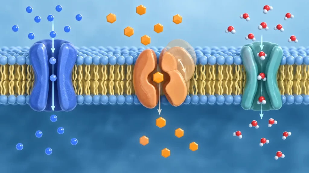
Facilitated diffusion is passive movement across a cell membrane with help from a transport protein. The word facilitated means that a membrane protein supplies a route that the substance cannot use efficiently through the lipid bilayer alone. Diffusion means that the net movement follows a favorable gradient rather than being driven uphill by the cell. A glucose transporter and an ion channel can both participate, although they work in different ways. Neither example means that the protein creates energy or that every molecule in the cell moves in one direction. The protein changes how readily a particular substance can cross; the existing gradient determines the direction of net flow.
This distinction matters in AP Biology because a drawing of a membrane can show a protein, an arrow, and a concentration difference without naming the process. You must infer whether the arrow is down a relevant gradient, whether the solute needs a channel or carrier, and whether another source of energy is coupled to the movement. A protein by itself does not prove active transport. Likewise, the absence of an ATP symbol does not prove passive transport: secondary active transport can use an ion gradient built by another process. The strongest explanation identifies the transported substance, its gradient, the protein’s role, and the energy relationship.
The familiar phrase “high to low concentration” is a useful starting point for an uncharged solute, but it is incomplete for ions. An ion experiences both a concentration difference and an electrical difference across the membrane. Together they form an electrochemical gradient. A positively charged ion can be pulled toward the negatively charged side even when its concentration difference points the other way. Thus, a question about sodium or potassium requires more than counting particles. Ask about membrane voltage as well as concentration, and state assumptions if the voltage is not given.
Why a membrane needs selective routes
The plasma membrane consists largely of a phospholipid bilayer. Its water-facing surfaces are hydrophilic, while the interior made of fatty-acid tails is relatively hydrophobic. This architecture separates intracellular and extracellular fluids and helps the cell maintain different conditions on either side. Small nonpolar molecules such as oxygen can cross the lipid region comparatively readily. Charged particles and many large or strongly polar molecules face a much larger barrier. That is why a pathway made by a membrane protein can have a profound effect on transport without changing the basic direction that diffusion favors.
A membrane is selectively permeable, not perfectly sealed. Its permeability depends on the substance, the bilayer, and the proteins present. For example, an ion channel offers a water-friendly route for particular ions. A glucose carrier recognizes glucose and alternates its access from one side to the other. Some water can pass through a bilayer, but aquaporins can make water movement much faster in membranes where they are present. No single rule such as “all polar molecules need channels” captures this variety. The useful question is how readily this substance crosses this membrane under these conditions.
The structure of a protein is also part of selectivity. A channel’s pore may favor a particular size and charge, while a carrier has a binding site and a cycle of shape changes. Proteins are not interchangeable holes. They can be gated, regulated, present in different numbers, or concentrated in a particular membrane region. A cell can therefore change permeability even when the outside concentration stays the same. This is one reason diagrams that show only colored dots on either side of a line can be misleading: the kinds and states of proteins in the line are essential information.
If membrane structure is new to you, revisit phospholipids and other biological macromolecules before memorizing transport vocabulary. The amphipathic nature of phospholipids explains why the bilayer forms in water and why its interior resists ions. That structural explanation gives the transport terms a cause rather than leaving them as disconnected definitions. It also prepares you to explain what would change if membrane composition or the abundance of a transporter changed.
What a gradient actually tells you
A concentration gradient is a difference in concentration between locations. Individual molecules move in random directions, but if more of a substance is on one side of a permeable boundary, more molecules tend to cross from that side per unit time than in the reverse direction. That imbalance is net movement. There can still be movement both ways, even while the net flow is strongly in one direction. At dynamic equilibrium, crossings continue but their average rates balance. “Equilibrium” does not mean every molecule has stopped or every cellular substance has the same concentration everywhere.
For an uncharged solute using one facilitated-diffusion carrier, net movement normally proceeds from higher free-solute concentration toward lower free-solute concentration. The carrier can speed exchange, but it cannot by itself maintain net movement against that gradient. If concentrations reverse, the possible net direction reverses too, assuming the carrier can function in both directions and no other process intervenes. In a living cell, metabolism, storage, and transport by other proteins can continually change concentrations. Therefore a classroom statement that glucose “always enters” is too broad; the direction depends on the specific membrane, transporter, and conditions.
For ions, use the electrochemical gradient. Suppose sodium is more concentrated outside a cell and the cell interior is negative relative to the outside. Both the concentration difference and electrical attraction favor Na+ entry through an open sodium channel. Suppose instead a cation has a concentration difference favoring exit while the electrical difference favors entry. You cannot decide net direction from the concentration numbers alone. You would need the membrane potential and enough information about that ion’s equilibrium tendency. AP-level reasoning usually asks you to identify both components, not calculate a full electrochemical potential.
Water movement is described as osmosis: net movement of water across a selectively permeable membrane in response to differences in the effective concentration of solutes that cannot freely follow it. Aquaporins are channels that can accelerate this movement. A cell in a solution with more nonpenetrating solute outside may lose water; in the opposite case it may gain water. Do not say water moves “to the side with more water” without identifying the relevant solutes and permeability. And do not assume that every solute shown in an osmosis diagram is impermeable. The problem’s assumptions determine the prediction.
Channel proteins: a route through the membrane
A channel protein forms a passage that allows an appropriate substance to cross the membrane. Many ion channels have a selective pore and may open or close in response to a stimulus. An open channel does not push an ion in a chosen direction. The electrochemical gradient supplies the driving force, and the channel offers a route. An open potassium channel may allow K+ to move outward in one set of conditions and inward in another. Potassium is K+, not K−; getting the charge right matters when explaining electrical forces across a membrane.
Gating helps a cell control when a route is available. Voltage-gated channels respond to changes in membrane potential; ligand-gated channels respond to binding of a signaling molecule. Other channels are influenced by mechanical forces or cell-specific signals. A channel that is closed may have the right selectivity yet carry little or no current at that moment. A channel that opens can rapidly change movement, but the net direction still depends on the gradient. Thus “gated” describes regulation of access, while “passive” describes the energetic relationship of the moving solute to its gradient.
Aquaporins are a useful channel example because water and ions should not be lumped together. Aquaporins provide selective water pathways; their structure helps exclude charged particles from the water route. Water movement through an aquaporin remains passive. The cell is not spending ATP at the aquaporin to force each water molecule across. Changes in the number or location of aquaporins can alter how quickly water crosses a membrane, but they do not change the thermodynamic direction that the water gradient favors. In a question, distinguish the protein’s effect on rate from the solution’s effect on net direction.
Channels are often described as fast relative to carriers because an open pore can let many particles pass without a complete binding-and-release cycle for every particle. That contrast is a tendency, not an absolute ranking of every biological protein. The actual flux depends on channel opening, selectivity, the magnitude of the gradient, and the number of functional proteins. A question that says “more channels were inserted” may predict greater transport capacity under the same gradient, but it should not be answered as though the concentration gradient itself grew stronger.
Carrier proteins: binding and changing shape
A carrier binds a particular solute, changes conformation, and releases it on the other side of the membrane. The transport cycle alternates which side can access the binding site. It does not leave a permanently open tunnel like a channel. A carrier can be highly selective, and its shape changes place a limit on how many molecules it can move in a given time. Those observations help explain why transport rate may rise with substrate concentration and then approach a plateau when available carriers are working near their capacity.
GLUT family proteins are familiar examples of glucose transporters that can mediate facilitated diffusion. A GLUT transporter does not consume ATP directly to carry each glucose molecule down a concentration gradient. However, the cell’s metabolism can remove or modify glucose after entry, helping maintain a difference in free glucose. Other glucose-transport systems, such as sodium-coupled glucose uptake in certain tissues, use a different mechanism: the sodium gradient drives coupled movement and can move glucose against its own gradient. Calling every glucose entry route “facilitated diffusion” erases this important distinction.
The distinction applies to amino acids too. Some transporters exchange or cotransport amino acids with ions, and the overall mechanism may depend on an ion gradient. Do not classify all amino-acid uptake as passive simply because a protein is involved. Identify what else moves, whether the solute travels with or against its own gradient, and how the driving gradient is maintained. A single membrane protein can be part of a larger energetic system. To classify the transport, analyze the whole coupled event described in the question rather than the name of the molecule alone.
Carrier-mediated transport can show specificity and competition. If two similar molecules use the same binding site, adding more of one may reduce the other’s transport, depending on the carrier’s properties. This is a reason to expect different rate patterns from transport through the lipid bilayer alone. Nevertheless, a plateau in a graph is not by itself proof of exactly which protein is present. A careful interpretation says that the graph is consistent with limited transport capacity, then looks for additional evidence about binding, inhibition, or protein abundance.
Facilitated diffusion, simple diffusion and active transport
Simple diffusion and facilitated diffusion are both passive in the sense that the transported substance moves down its relevant gradient. The difference is the route. In simple diffusion, the substance crosses the lipid bilayer without a dedicated transport protein; in facilitated diffusion, a channel or carrier helps it cross. Protein assistance often makes movement faster or more selective. It does not change a downhill trip into an uphill one. When comparing diagrams, focus on the pathway and the gradient rather than assuming “protein equals active.”
Active transport moves a substance against its own electrochemical gradient, directly or indirectly using an energy source. In primary active transport, a transporter can use ATP hydrolysis. The sodium-potassium pump, for example, moves Na+ and K+ in directions that help maintain their gradients and consumes ATP. It is not a channel for facilitated diffusion. Those gradients can later power passive ion movement through channels or coupled transport through other carriers. The pump and a channel may act in the same cell, but they perform different energetic jobs.
Secondary active transport uses an existing ion gradient as an energy source. A cotransporter may let sodium move down its favorable gradient while carrying another substance uphill. The cotransporter may not split ATP itself, yet maintaining the sodium gradient often depends on ATP-using pumps elsewhere in the cell. “No ATP attached to this protein” is therefore not enough to label a diagram passive. Trace each solute’s direction relative to its own gradient and ask whether one downhill movement is coupled to another uphill movement. This is the central reasoning distinction.
Bulk transport is different again. Endocytosis brings material into a cell by forming a vesicle; it is not a solute slipping through a membrane protein. If your diagram shows a membrane folding around material, the relevant mechanism is vesicular transport, not facilitated diffusion. The Sly Academy overview of endocytosis and bulk uptake provides a useful comparison. Recognizing the scale and physical route of the material helps prevent a common classification error in microscopy images and schematic drawings.
A comparison you can reason through
Imagine three identical artificial membranes, each separating two solutions with more of an uncharged solute on the left. The first membrane has no path for that solute. The second contains a specific passive carrier. The third contains a pump supplied with an energy source. In the first setup, crossing may be negligible if the solute cannot pass the bilayer. In the second, net movement can occur left to right while the gradient remains favorable. In the third, the pump could, if designed for this solute, maintain a concentration difference or move material against it. The arrow in a diagram is meaningful only alongside the route and energy information.
Now remove the energy supply from the third membrane. If the pump depends on that energy, its uphill transport cannot continue in the same way. The passive carrier in the second membrane, however, can still work while a gradient remains. That does not mean the whole living cell needs no energy. It means that this particular carrier step does not require direct energy input to move its substrate downhill. Separating the energetics of one transport step from the energy needs of the entire cell makes many exam questions much easier.
Try another variation: add ten times as many passive carriers to the second membrane while keeping the same initial concentrations. The maximum possible transport rate may rise because more routes are available. The favorable direction does not reverse, and the initial gradient is not made steeper by the carriers alone. As solute redistributes, the gradient may diminish unless another process replenishes it. This is why statements about a transport protein’s effect on rate should be distinguished from statements about the equilibrium state or net direction.
Rate, saturation and dynamic equilibrium
At low substrate concentration, adding more substrate may increase the rate of carrier-mediated transport because more binding sites are occupied during a given interval. At high concentration, a finite number of carriers can become the limiting factor, so the rate approaches a maximum under those conditions. A graph with a curve that levels off is often used to illustrate saturation. Do not interpret the plateau as proof that molecules have stopped moving. It says the available carriers have a finite throughput, assuming the experimental design actually isolates that process.
Opening additional channels can also raise flux when a suitable gradient exists. The relationship is not identical to a carrier binding curve, because a channel’s pore can support passage of many particles while open. But channel movement can still be limited by the number of open channels, their conductance, and the electrochemical driving force. If a question asks why a rate changes after a signaling molecule binds, consider whether the signal changed gating, transporter number, or the gradient. One observed rate change can have several possible mechanisms.
At dynamic equilibrium for an uncharged substance across a simple permeable boundary, movements continue in both directions but net movement averages to zero. In a living cell, true equilibrium for every substance is unusual because cells continually metabolize molecules, pump ions, exchange materials, and respond to their surroundings. A glucose carrier may keep moving glucose molecules both ways while metabolism keeps free intracellular glucose relatively low. Therefore a biological graph may show a sustained net flow without contradicting the passive nature of the carrier step. The wider system maintains the conditions.
Ion channels and electrical signaling
In excitable cells, opening ion channels can rapidly change membrane potential. That does not mean the channel “manufactures” an action potential or pumps ions against their gradients. Sodium and potassium gradients, established and maintained by other processes, provide the potential for movement. Channel opening changes permeability, and ion flow changes electrical conditions. Because the electrical conditions then change, the driving force for an ion can change during the event. A simplified AP Biology diagram often freezes one moment of this dynamic process; describe what the diagram actually shows without assuming a permanent direction.
Suppose an open potassium channel is shown with much more K+ inside than outside. Concentration alone favors outward movement, but the negative cell interior favors K+ entry. The net direction depends on the balance of those effects. If the problem gives only concentrations, say the concentration component points outward and acknowledge the missing voltage. If it gives a membrane potential as well, use both. This is more scientifically sound than claiming that potassium must always leave through a channel or treating positive potassium as a negative ion.
The sodium-potassium pump deserves its own sentence in an exam response because students often confuse it with an ion channel. It uses ATP to move sodium outward and potassium inward, helping preserve the gradients. A separate sodium channel or potassium channel allows passive movement when open. The pump is analogous to charging a battery, while a channel can provide a circuit through which stored electrochemical energy is released. The analogy is imperfect—the cell is not a simple battery—but it clarifies why the pump and the passive channel should not be placed in the same transport category.
Worked AP Biology practice questions
Question 1: classify a transport diagram
A diagram shows a polar, uncharged molecule more concentrated outside a cell than inside. It binds to a membrane protein, the protein changes shape, and the molecule is released inside. No other solute moves with it, and there is no energy source attached to the step. The best classification is facilitated diffusion by a carrier. The molecule is moving down its concentration gradient, and the protein provides a selective path. The shape change does not make the process active; many passive carriers change conformation. To earn a strong explanation, name both the direction relative to the gradient and the carrier mechanism.
Question 2: change transporter abundance
Researchers double the number of functional carriers in an otherwise comparable membrane and measure initial uptake while the concentration difference is held constant. A reasonable prediction is that uptake capacity can increase, especially when the original carriers limited the rate. The prediction is not that the solute will now move against its gradient. It is also not that every condition will show precisely double the measured rate: other steps, available solute, and the experimental interval may limit flux. State the controlled condition and the mechanism behind your prediction rather than repeating a memorized slogan about more proteins.
Question 3: compare a pump and a channel
A cell uses ATP while moving sodium outward even though sodium would tend to enter through an open sodium channel. The ATP-linked outward step is primary active transport if a pump directly couples ATP hydrolysis to Na+ movement. The inward channel step is passive ion movement down a favorable electrochemical gradient, provided the given electrical and concentration conditions favor entry. These processes can occur in the same cell and at different times. A response that says both are “facilitated diffusion because proteins are involved” ignores the role of energy and reverses the classification.
Question 4: explain an ambiguous ion arrow
Imagine a graph reports higher K+ concentration inside a cell, and a diagram shows an open K+ channel but gives no membrane potential. The concentration component favors movement outward. The electrical component is unknown. The most defensible answer is conditional: if the combined electrochemical gradient favors exit, net K+ flow will be outward; if the electrical attraction to the interior is strong enough, that conclusion could change. This response does not evade the problem. It identifies exactly which missing measurement would resolve it and avoids treating concentration as the only physical influence on a charged particle.
Question 5: water and a new aquaporin
Two cells are placed in the same external solution. One has more functional aquaporins in its membrane. If the solution and other conditions create a water gradient, that cell may change volume faster because its water permeability is higher. The extra aquaporins do not necessarily change the final direction of net water movement or the external solution’s tonicity. To answer correctly, separate rate from direction and remember that osmosis depends on solute behavior as well as protein number. If the diagram does not tell you whether the solutes can cross, make that uncertainty explicit.
How to interpret evidence rather than memorize a label
When given a transport graph, read the axes and units before naming a mechanism. Is the vertical axis amount transported, rate, or concentration inside the cell? Is the horizontal axis time, external concentration, or ATP availability? A declining net uptake rate over time could reflect a weakening gradient rather than loss of proteins. A plateau as concentration rises may suggest finite transporter capacity. If a drug blocks ATP production and uptake falls, the observed effect could be direct or indirect because energy-dependent pumps may maintain the gradient used by another transporter. Design and controls matter.
A useful experimental comparison holds several conditions constant and changes one plausible cause. To test whether a particular protein facilitates uptake, compare otherwise similar cells with and without that protein, while measuring the relevant concentration gradient and checking cell health. If uptake increases when the protein is present and remains aligned with the gradient, that is evidence consistent with a passive route. It is not absolute proof from one graph; other membrane changes could contribute. Good AP Biology explanations connect a prediction to the cell mechanism and identify a control that makes the inference stronger.
Another evidence question concerns specificity. If adding a similar molecule reduces transport of the original substrate, competition for a carrier’s binding site is one possibility. But the added molecule might also change membrane conditions or cell metabolism. A careful answer says what observation fits the model and what comparison would distinguish alternatives. Scientific reasoning is not just matching the word “carrier” to a curve. It is using structure, gradient, and controlled evidence to decide which explanation best fits the data.
Common misconceptions and quick corrections
“Facilitated diffusion uses ATP because a protein is working” is incorrect. A protein can change shape or open a gate without directly using ATP for the downhill movement under discussion. “All protein-mediated transport is facilitated diffusion” is also wrong, because pumps and coupled transporters can move a solute uphill. “All solutes move until their concentrations are equal” overlooks metabolism, electrical forces, and ongoing transport. “A closed channel proves there is no gradient” confuses availability of a route with the force that would drive movement if the route opened.
“Osmosis is always the same as facilitated diffusion” is too broad. Water can cross some lipid bilayers directly, and aquaporins can greatly accelerate water passage; the central term osmosis describes the net water movement under an appropriate gradient. “Potassium is K−” is a chemical error: potassium ions in these examples are K+. “The sodium-potassium pump is an ion channel” misses that the pump uses ATP and performs uphill transport. Each correction has the same underlying lesson: specify the substance, the route, the relevant gradient, and the energy source.
A practical study method
Draw a bilayer and annotate four separate routes: direct passage through the lipid, an open channel, a cycling carrier, and an ATP-driven pump. For each route, write one sentence saying what passes, which way net movement would go under stated conditions, and what energy relationship applies. Then redraw the diagram with the concentration difference reversed. Ask which arrows reverse and which processes require an additional energy source to keep working. This exercise tests understanding more effectively than copying a list of definitions because it forces you to use the conditions in the question.
Make a second version with Na+ and K+ and add a membrane voltage. For the ions, write “electrochemical gradient” rather than only “concentration gradient.” For glucose, compare a downhill GLUT route with sodium-coupled uptake and identify the hidden role of a pump that maintains the sodium gradient. For water, add aquaporins and distinguish speed of volume change from its direction. Finally, explain each sketch aloud without using the word “because” only once. A full causal explanation usually needs membrane structure, a transport protein, and a driving force.
If you need to reconnect this topic to the larger biological picture, revisit the basic structure and function of a cell. Membrane transport is not an isolated vocabulary unit; it supports nutrient uptake, waste removal, signaling, and homeostasis. The College Board AP Biology course overview places membrane structure and function within a broader study of how biological systems maintain organized conditions. Use that official framework for course scope rather than assuming a particular old article’s term list is the exam blueprint.
Conclusion and authoritative references
Facilitated diffusion is a precise combination of pathway and direction: a channel or carrier helps a substance cross a membrane while the net movement follows a favorable gradient. Channels form selective passages; carriers bind substrates and change shape. Both can alter permeability and rate without serving as an ATP-driven pump. For ions, consider the electrical and concentration components together. For glucose, distinguish a downhill carrier from sodium-coupled uptake. For water, distinguish the aquaporin route from the osmotic gradient. When you can explain those contrasts with a diagram or experimental result, you understand the concept rather than merely recalling its name.
For a textbook treatment, consult OpenStax Biology 2e on passive transport and its companion chapter on active transport. These sources support the distinctions between downhill transport, pumps, and coupled movement. The College Board AP Biology exam overview is the official place to check current assessment information. The worked scenarios above are original teaching examples with stated assumptions, not accounts of a particular research study or a claim that every membrane behaves identically.