
Giant cell tumor of bone is rare and usually noncancerous. This guide explains its symptoms, diagnosis, treatment choices and follow-up, with practical questions for specialist care.
What a giant cell tumor of bone means
Giant cell tumor of bone, often shortened to GCTB, is a rare tumor that starts in bone. Most giant cell tumors are benign, meaning noncancerous, although malignant forms exist. The diagnosis is seen most often in young and middle-aged adults and commonly involves bone near a joint. It is useful to keep the complete name when discussing the diagnosis: “giant cell tumor” without its tissue of origin can be ambiguous. The word tumor alone does not establish that someone has cancer. [1]
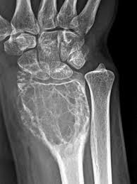
GCTB usually develops after skeletal growth is complete, often in young or middle-aged adults. Common sites include the ends of the thighbone and shinbone near the knee, and the wrist. It can also affect other bones, including the pelvis and sacrum. Most cases are noncancerous but locally aggressive. Rare lung spread can occur even when the tumor retains its conventional, nonmalignant microscopic appearance; metastasis does not automatically establish malignant transformation. Persistent local pain, swelling, stiffness or reduced joint movement may be the first signs. Some people notice a lump without much pain. Symptoms alone cannot identify the tumor type. [2]
Specialists also use the category intermediate for a bone giant-cell tumor that is benign but locally aggressive. This terminology describes behavior rather than the amount of pain a person feels. Ask the team to explain the wording in your own report, particularly if “intermediate,” “aggressive” and “malignant” appear in different documents. [3]
A similarly named condition, tenosynovial giant cell tumor or TGCT, starts in tissue lining a joint or a tendon sheath. Older terms include pigmented villonodular synovitis and giant cell tumor of tendon sheath. TGCT and GCTB are different diseases, even though both can occur close to joints and contain large cells under the microscope. The location of a lump is therefore not enough to distinguish them. Check the exact wording of the pathology report when reading treatment information or discussing a referral. Research about TGCT, and medicines approved specifically for TGCT, should not be transferred to GCTB. This article concerns the bone tumor. [4]
What happens inside the tumor
The large cells that give GCTB its name are not the whole story. A landmark genetic study identified changes in H3F3A, a gene involved in a histone protein that helps organize DNA. These changes were found in the tumor’s stromal cells rather than in the recruited giant cells. They help explain why identifying the underlying tumor cell population matters. A somatic change develops in affected cells; finding it in a tumor does not by itself show that a person inherited a condition or will pass it to a child. A laboratory finding also cannot predict an individual’s future with certainty. Ask whether a test was performed on tumor tissue, what it supports and whether any separate genetic counseling is actually indicated. [5]
Stromal tumor cells can produce RANK ligand, usually written as RANKL. This signal helps recruit and activate osteoclasts, cells whose normal job includes breaking down bone during remodeling. In GCTB, the interaction between the tumor cells and these bone-resorbing cells contributes to local bone loss. Laboratory research has demonstrated strong RANKL expression in GCTB, providing a biological reason to target that pathway. It does not mean that every bone lesion expressing RANKL should receive the same medicine. A treatment target is one part of the evidence needed for clinical care; diagnosis, clinical studies, product authorization and the person’s circumstances remain essential. Understanding the mechanism also helps explain why reducing bone destruction is not necessarily the same as eliminating every tumor cell. [6]
Symptoms that need prompt or emergency attention
Sudden severe pain, a new deformity or inability to put weight through a leg can indicate a fracture. Seek urgent medical assessment rather than testing the limb repeatedly to see whether it improves. Avoid loading a suspected broken leg while arranging help. An open wound over a possible fracture, an abnormally positioned limb or a cold or discolored foot warrants emergency care. These are general fracture warning signs, not a way to diagnose GCTB. When a known bone lesion is present, tell the emergency team and bring available treatment information. Pain that has changed abruptly should not wait for a routine tumor-clinic appointment. [7]
For a lesion involving the spine or sacrum, new nerve symptoms require particular care. Seek emergency assessment for new loss of bladder or bowel control, difficulty starting or passing urine, numbness around the genitals or buttocks, or worsening leg weakness, especially with back pain. Pressure on the lower spinal nerves can interfere with movement, sensation and bladder or bowel function. Early assessment matters because the consequences can be lasting. These symptoms have several possible causes and do not prove that a tumor has progressed. Their urgency comes from the possible nerve problem. Do not wait for several warning signs to appear together or assume that a scheduled scan will be soon enough. [8]
How the diagnosis is established
An X-ray is usually the first imaging test for a suspected primary bone tumor. It shows where bone has been lost or altered and helps the team decide what to examine next. For an uncertain or aggressive-looking lesion, MRI or CT can clarify its extent and support biopsy or surgical planning. MRI is especially useful for mapping the relationship to surrounding tissues, while CT can provide additional bone detail. The American College of Radiology bases imaging choices on the clinical situation rather than recommending every scan for every patient. An alarming phrase in a radiology report describes an appearance that needs investigation; it is not a final tissue diagnosis. Ask what the next scan is intended to resolve and how its result will change the plan. [9]
A biopsy removes a sample for examination by a pathologist. It may use a needle or require a small operation, depending on the lesion and the information needed. For a suspected primary bone tumor, the biopsy should be planned with the orthopedic oncology team before definitive surgery. The route matters because its position can influence a later operation. An apparently convenient approach may interfere with options for removing or reconstructing the affected area. Imaging and pathology must be considered together, and an uncertain sample may need further review. Asking for specialist planning is therefore part of obtaining a reliable diagnosis and preserving treatment choices. It is reasonable to ask who will perform the biopsy and who will interpret the tissue. [10]
Pathologists may use a stain for the H3.3 G34W protein change to help identify GCTB. Research has shown that this can distinguish many GCTBs from other lesions that also contain giant cells. However, a negative result does not automatically rule out GCTB: some tumors carry a different alteration or do not show the expected staining. Conversely, a positive marker is interpreted within the entire specimen and clinical picture. Giant-cell-rich lesions include aneurysmal bone cyst, chondroblastoma and some malignant bone tumors. The team needs the specific diagnosis rather than the descriptive phrase “giant cells present.” If the findings are unusual, review by a bone-tumor pathologist can clarify what additional tissue studies or molecular tests are appropriate. [11]
Blood tests can help investigate alternative explanations and assess general health, but they do not replace imaging and tissue assessment for a suspected bone tumor. One relevant alternative is excessive parathyroid hormone activity, which can remove calcium from bone and weaken it. Clinicians interpret calcium together with parathyroid hormone and other findings when investigating hyperparathyroidism. An abnormal calcium result has more than one possible explanation, and a result within the laboratory range needs clinical context. Ask what a requested blood test is checking: an alternative diagnosis, kidney function, readiness for a procedure or medicine safety. Those purposes are different. Do not assume that a normal routine blood panel has ruled out a bone lesion that still needs specialist assessment. [12]
A “brown tumor” caused by hyperparathyroidism is one important giant-cell-rich mimic. Identifying this alternative matters because treating the hormone disorder and treating true GCTB involve different plans. [13]
Choosing treatment while protecting bone and joint function
Surgery is the usual treatment for resectable GCTB. The central choice is often between curettage, which removes tumor from inside the bone, and resection, which removes the involved segment. Curettage can preserve more of the natural joint and surrounding structures, while resection generally offers stronger local control at the cost of a larger reconstruction and possible functional loss. The 2024 British Sarcoma Group consensus recommends management by a specialist bone-tumor multidisciplinary team. The decision depends on the tumor’s extent, the condition of the joint, soft-tissue involvement and the consequences of each operation. Ask the surgeon to explain both the planned procedure and the alternative, including why the expected balance is appropriate for your particular anatomy. [13]
During extended curettage, the surgeon opens a route into the bone, removes the visible tumor and carefully treats the cavity. A high-speed burr can help clear the cavity margins. Local adjuncts and a filler may also be used, but practices vary. A retrospective series of more than two hundred patients examined combinations of burring, bone cement and other local measures, supporting careful local clearance while illustrating how techniques differ between treatment groups. Such a study does not prove that one combination is best for everyone today. Ask which steps your operation includes, why they are suitable near your joint and what the surgeon considers the main remaining risk. These are operating-room techniques, not substances or procedures to use outside specialist care. [14]
After curettage, the remaining space may be supported with bone graft, cement or a combination. When a lesion lies close to a joint, the remaining layer of bone beneath the cartilage is particularly important. Long-term research on knee GCTB treated with curettage and cement found that arthritis can develop, although many changes were mild. That finding helps frame follow-up: successful tumor control and joint health are related but separate outcomes. Later discomfort may come from arthritis or a mechanical problem rather than recurrence, and should be assessed instead of guessed at. Ask how the reconstruction protects the joint surface, whether fixation is needed and what symptoms should prompt review of the repair. [15]
A pathologic fracture occurs when disease has weakened a bone enough for it to break. It can make surgery more complicated, but it does not automatically mean that the whole affected segment must be removed. A comparative study of GCTB with fractures found different tradeoffs after curettage and resection, including local control, complications and function. The presence of a soft-tissue component, a complex fracture or inability to restore a stable structure can influence the choice. The study was retrospective, so its results should guide discussion rather than serve as a rule for every fracture. Ask whether the joint surface remains reconstructable, how stability will be achieved and how the fracture changes the rehabilitation plan. [16]
For a tumor at the wrist, preserving movement is only one part of preserving useful hand function. Some extensive lesions require removal and reconstruction of the distal radius, and selected cases may involve fusion of the wrist. Fusion changes movement but can provide a stable, usable joint. A small long-term study compared patient-reported results after joint-preserving treatment and more extensive wrist reconstruction. Its findings emphasize that a larger operation is not automatically a worse functional result when the joint surface is already involved. They do not establish one preferred operation for all wrist tumors. Discuss grip, lifting, dominant-hand tasks, work demands and the activities that matter most to you alongside recurrence risk and the likelihood of further surgery. [17]
When denosumab may be considered
In the United States, Xgeva, a denosumab product, is approved for adults and skeletally mature adolescents with GCTB that is unresectable or for which surgical resection is likely to cause severe morbidity. This June 2026 label does not make denosumab routine treatment for every GCTB. It is not a reason to substitute Prolia or another product. Important risks include severe low blood calcium, osteonecrosis of the jaw and atypical thighbone fractures. Low calcium must be corrected before treatment; severe kidney impairment increases this risk. Calcium monitoring, a dental assessment and a clinician-directed supplement plan are important; report jaw symptoms or new thigh or groin pain. Invasive dental work needs coordination with the prescribing team. High calcium and multiple vertebral fractures can occur after treatment stops. Denosumab can harm a developing pregnancy, so pregnancy testing and contraception counseling are needed when applicable. Discuss breastfeeding too. Do not stop, restart or change treatment independently; the team must plan both treatment and follow-up after discontinuation. [18]
The large international phase 2 denosumab study included people whose tumors could not be removed surgically and people whose planned operation was expected to cause major harm. It documented tumor control and, in some patients, avoidance or reduction of the originally planned surgery. Earlier reports from the same trial examined surgical downstaging. These are reasons a specialist may consider treatment when preserving a joint or avoiding a highly disabling operation is important. However, the trial was open-label and did not randomly compare denosumab with an untreated control group. It does not establish a universal cure rate, the best duration for every patient or a guarantee that surgery will become unnecessary. The relevant question is what specific benefit the team expects in your case and how it will be measured. [19]
Blocking bone-resorbing cell activity does not necessarily eliminate all tumor cells. Laboratory work comparing denosumab with another bone-targeting medicine found that denosumab did not eliminate the growth of the neoplastic stromal cells in the cultures studied. This is laboratory evidence, not a prediction that every patient will relapse. It does explain why an improvement in bone destruction cannot simply be called permanent eradication. The treatment team interprets imaging alongside examination and, when relevant, surgical pathology. Ask what a reported response means: less pain, less bone destruction, stable disease or a change in the operation that can be offered. These outcomes are useful but not interchangeable, and the planned assessment should match the intended treatment goal. [20]
Using denosumab before curettage needs a careful discussion of uncertainty. A retrospective study found more local recurrence in patients who received denosumab before surgery than in historical comparison patients treated without it. The authors also noted important differences between the groups and could not establish causation. More difficult tumors may be more likely to receive the medicine in the first place. The finding therefore raises a concern; it does not prove that denosumab always causes recurrence or should never be used before surgery. Ask why pretreatment is being recommended, whether it changes the planned operation, how the surgeon will account for the treated tumor and what surveillance will follow. A good rationale should describe both the expected benefit and the unresolved tradeoff. [21]
There is no single established treatment duration that suits every unresectable GCTB. A small multicenter study published in 2025 followed patients after denosumab ended: some remained stable, while others developed progression. Because only a small selected group was studied retrospectively, the results cannot identify a reliably safe stopping point. They also argue against claiming that recurrence after stopping is inevitable. Decisions about continuing, spacing or ending treatment belong with the specialist team and should consider disease control, adverse effects and available alternatives. Before a planned change, clarify which symptoms to report, which imaging and blood tests are needed and who will arrange them. A favorable scan is part of that decision, not permission to miss treatment or monitoring. [22]
Less common treatments for difficult locations
Selected tumors in the sacrum or pelvis may be considered for arterial embolization. An interventional specialist deliberately blocks vessels supplying the tumor, sometimes to help another treatment or reduce surgical bleeding. An older long-term series of sacral GCTB showed that some patients had durable symptom and imaging improvement, but late recurrence and serious procedural risk were also reported. These results support discussion in a specialist team, not routine embolization for every bone tumor. The anatomy matters because nearby nerves and normal tissues also depend on their blood supply. Ask whether the intended goal is preparation for surgery, longer-term control or symptom relief, whether repeat procedures might be needed and what other options are available. Ongoing monitoring remains necessary even after a good early response. [23]
Radiotherapy has been studied in selected unresectable, recurrent or incompletely removed GCTB. A retrospective Rare Cancer Network series reported local control in many treated patients, but it involved selected cases and older treatment eras. It cannot establish that radiation is the best first choice today or eliminate concern about late effects. Modern planning can shape the treated area, yet the balance still depends on nearby nerves, joints and other tissues, previous treatment and the available surgical or medical alternatives. If radiation is proposed, ask why those alternatives are unsuitable, what benefit is expected and how long-term effects will be monitored. A specialist recommendation should explain the particular difficulty that radiation is intended to address. [24]
AAOS guidance describes radiotherapy as a rare option when surgery is difficult. Its possible late risks, including a radiation-related malignancy, require discussion. It is not a routine replacement for surgery in readily operable GCTB. [2]
Follow-up, recurrence and lung checks
Follow-up checks the original tumor site and, when appropriate, the lungs. Local examination and imaging also help assess the reconstruction. A study of lung surveillance found that local recurrence was associated with a higher likelihood of lung metastasis and that CT detected lung lesions more sensitively than chest radiography in that cohort. This supports reconsidering the chest-imaging plan when local disease returns. It does not supply one scan timetable for everyone. The treating center chooses the imaging method and interval using the tumor’s course, previous treatment and practical considerations, including radiation exposure. Ask for a written plan naming the next appointment, the tests due and the clinician who will review the results. Report new problems between visits instead of waiting for surveillance to find them. [25]
A 2026 single-center study also found an association between local recurrence and pulmonary disease, while emphasizing how uncommon lung metastasis was in the population examined. The authors proposed a more selective chest-imaging approach. That proposal is not a universal standard: it came from retrospective data, and not every patient in the overall cohort had chest imaging. Different centers may reasonably assess the evidence differently. If your schedule changes, ask whether this reflects your own tumor history, a new finding or the center’s protocol. Do not cancel a recommended scan because another study used a different approach. [26]
Local recurrence means that GCTB has returned at or near the treated bone. It is not automatically malignant transformation, and it does not mean that all useful treatment options are exhausted. A study of recurrent tumors in long bones found that selected patients could undergo further curettage, while other situations required resection. The amount of remaining bone, the condition of the joint and earlier reconstructions affect what is feasible. The decision also needs renewed imaging and specialist review rather than an assumption that the first operation should simply be repeated. Ask what has changed since the original treatment, what the proposed operation can preserve and what further treatment would remain possible if another recurrence occurred. These are individualized decisions, not a verdict on anything the patient did. [27]
GCTB can rarely form deposits in the lungs without showing the high-grade sarcoma features of malignant GCTB. The behavior of these deposits varies. A small study of patients whose lung lesions were initially observed found that some remained stable, while others progressed and needed treatment. Carefully selected observation can therefore be an active management choice, with planned imaging and a threshold for intervention. Other patients may need lung surgery or medical treatment. A new lung nodule also requires interpretation; its presence on a scan does not automatically identify its cause. Ask what supports the diagnosis, whether the finding has changed and why the team recommends surveillance or treatment. Breathlessness or chest symptoms deserve clinical assessment and should not be attributed to a scan finding without review. [28]
Malignant GCTB is different from conventional GCTB behaving aggressively in its original bone or forming nonmalignant-appearing lung deposits. Pathologists identify a high-grade sarcoma component. It may be present at the initial diagnosis or arise later at a previously treated site. A small single-center study of secondary malignancy illustrates that this can occur without previous radiotherapy, but such a selected series cannot give a reliable personal probability. An unexpected change in symptoms or imaging may lead to renewed biopsy and pathology review. It does not establish malignancy on its own. If the diagnosis changes, ask the team to explain the evidence and how the treatment pathway changes. The current specimen and the earlier diagnosis may both need review. [29]
Chemotherapy is not standard treatment for conventional GCTB. If high-grade malignant transformation is confirmed, the specialist team considers a sarcoma treatment pathway rather than simply repeating the previous GCTB plan. [13]
Recovery and returning to daily activities
Rehabilitation should follow the operation and the strength of the reconstruction, not a generic GCTB exercise timetable. After a major reconstruction around the knee, for example, the surgeon and physiotherapist may prescribe specific weight-bearing limits, a brace and walking aids. Advice for one type of replacement should not be copied after a different procedure. Before leaving hospital, ask what you may load, how to use stairs and which movements are restricted. Have the team demonstrate exercises and check your technique. Progress is usually assessed against healing, stability and function rather than pain alone. Feeling better does not prove that a graft, repaired tendon or implant is ready for unrestricted activity. Ask who can reassess the plan if equipment, pain or the home layout makes it difficult to follow. [30]
Recovery is worth measuring in ways that reflect everyday life. A small cross-sectional study of adults with hip or knee tumor prostheses included people treated for bone sarcoma or GCTB and found continuing limitations in physical function and quality of life. Because it was a mixed group, its results are not a GCTB-specific prognosis or proof of what rehabilitation will achieve. They do show why a satisfactory scan should not end conversations about walking distance, fatigue, strength or participation. Tell the team which activities remain difficult and which improvements would matter most. Patient-reported function can reveal problems that are not obvious during a brief examination. Goals might need to include adaptations and assistive equipment as well as further improvement in movement. [31]
Living with a rare bone tumor can affect work, transport, caring responsibilities and confidence long after an operation. Support may include a clinical nurse specialist, physiotherapist, occupational therapist, dietitian or social worker, depending on the need and the local service. The Bone Cancer Research Trust describes holistic needs assessment as a way to discuss concerns beyond the tumor itself and create a support plan. It is reasonable to ask for similar help even if your hospital uses a different name. Worry before scans or after treatment ends is also worth raising. Peer support can help people feel less isolated, but another person’s operation, medicine response or follow-up schedule may not fit your case. Use shared experiences for support while keeping medical decisions with your own team. [32]
Persistent low mood, loss of interest, disrupted sleep or anxiety that interferes with ordinary life deserve assessment alongside physical symptoms. Long-term illness and depression can coexist, and depression remains treatable when someone has a physical condition. A clinician can consider pain, medicines, sleep and other contributors, and discuss counseling, medication or another appropriate approach. This does not mean the bone symptoms are imaginary or that emotional distress caused the tumor. Tell the mental-health clinician about ongoing treatment so care can be coordinated. Practical support and psychological care can address different problems at the same time. You do not need to wait until every scan or surgical issue is settled before asking for help with how you are coping. [33]
Research and making the next appointment useful
For an uncommon tumor, a specialist may discuss research participation when a suitable study is available. A trial listing is not a recommendation for your case, and an investigational treatment is not established care simply because early results appear encouraging. Ask about the study’s purpose, eligibility, alternatives, extra visits, tests, costs and the plan if treatment does not help. The research team should explain foreseeable risks and uncertainties clearly enough for an informed choice. Participation is voluntary, and declining should not prevent access to appropriate usual care. Discuss studies with the team before making travel or treatment arrangements. The National Cancer Institute’s trial-search guidance emphasizes comparing the proposed study with the other available options and checking the practical demands as well as the possible benefits. [34]
Bring your pathology wording, medication list and main questions. Ask what the treatment aims to achieve, what function it may change, which warning signs need urgent help and who coordinates follow-up. Request written instructions and a contact route for problems between visits. [35]
Frequently asked questions
Is GCTB cancer?
Usually not, but it can damage bone and rarely spread.
Is GCTB the same as TGCT?
No. TGCT begins in joint lining or tendon sheaths.
Is GCTB inherited?
A tumor genetic change does not automatically mean an inherited disorder.
Does every GCTB need bone resection?
No. Selected tumors can be treated with bone-preserving curettage.
Can GCTB return after treatment?
Yes. Follow-up remains important after treatment.
Can I stop denosumab when pain improves?
Do not stop treatment yourself; discuss changes and monitoring with your specialist.
Sources
- NCI: Definition of giant cell tumor
- AAOS: Giant cell tumor of bone
- NCI Thesaurus: Intermediate osteoclastic giant-cell-rich tumor of bone
- AAOS: Tenosynovial giant cell tumor
- Behjati and colleagues: Histone driver changes in bone tumors
- Primary study: RANKL expression in bone tumors
- NHS: Broken leg warning signs
- Oxford University Hospitals: Cauda equina syndrome awareness
- ACR: Suspected primary bone tumors, 2024 update
- NCI: Primary bone cancer and biopsy planning
- Amary and colleagues: H3.3 G34W diagnostic staining
- NIDDK: Primary hyperparathyroidism
- British Sarcoma Group: UK bone-tumor guidelines, 2024 consensus
- Balke and colleagues: Treatment outcomes in 214 GCTB cases
- Long-term study: Knee arthritis after GCTB curettage
- Van der Heijden and colleagues: Surgery for GCTB with fracture
- Kuruoglu and colleagues: Long-term wrist outcomes after GCTB treatment
- DailyMed: Xgeva US prescribing information, June 2026
- Chawla and colleagues: International phase 2 denosumab study
- Primary laboratory study: Denosumab effects on GCTB cells
- Errani and colleagues: Denosumab before curettage and recurrence
- DENO Research Group: Outcomes after denosumab discontinuation
- Lin and colleagues: Long-term embolization outcomes in sacral GCTB
- Bhatia and colleagues: Rare Cancer Network radiotherapy series
- Rosario and colleagues: Lung surveillance after GCTB treatment
- Kamel and colleagues: Chest imaging in GCTB, 2026
- Klenke and colleagues: Surgery for recurrent GCTB of long bones
- Tsukamoto and colleagues: Initial observation of GCTB lung metastases
- Single-center study: Secondary malignancy in GCTB
- RNOH: Rehabilitation after proximal tibial replacement
- Cross-sectional study: Function after hip or knee tumor prostheses
- Bone Cancer Research Trust: Living with GCTB
- NIMH: Chronic disease and depression
- NCI: Steps to find and evaluate a clinical trial
- AHRQ: Preparing questions for medical appointments