
Eye infection treatment depends on the cause and the part of the eye involved. This educational guide explains assessment, common treatments, safer supportive care, and warning signs. It cannot diagnose an individual eye problem. [1]
What eye infection treatment needs to address
Eye infection treatment starts by identifying what is affected and what is causing the problem. Conjunctivitis, often called pink eye, is inflammation of the membrane over the white of the eye and inside the eyelids. Viruses and bacteria can cause it, but so can allergens and irritants. That distinction matters because a red eye does not automatically need an antibiotic. The word infection also should not be used for every episode of itching, watering, or redness. A clinician considers the symptoms, exposure history, and examination together before recommending treatment. Some mild conjunctivitis can be managed with supportive care, while other causes need prescription medicine or urgent specialist assessment. The useful first question is therefore what the eye problem represents, rather than which bottle of drops to buy. [1]
The cornea is the transparent front surface of the eye. It bends incoming light, which then passes through the pupil and lens before reaching the retina. The retina converts light into signals that travel through the optic nerve to the brain. Tears also help the eye work normally. This arrangement explains why the location of a problem matters: redness at the surface and damage to a structure that focuses light are different concerns. Describing everything as an eye infection can hide those differences. Understanding the basic parts of the eye also makes an examination easier to follow. When a clinician discusses the cornea, pupil, or retina, they are identifying a particular structure, not simply using several names for the same area. [2]
Symptoms and warning signs that change the urgency
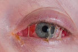
Conjunctivitis can cause redness, swollen lids, tears, crusting, burning, itching, and a feeling that something is in the eye. Discharge may be watery or thicker, and lashes can stick together after sleep. Viral cases often accompany a cold and may move from one eye to the other. Bacterial cases often produce thicker discharge, while allergy commonly causes marked itching in both eyes. These are patterns, not a dependable home diagnostic test. The same person can have several symptoms that seem to fit more than one category. Describe what happened and when it began rather than deciding the cause from discharge color alone. Pain, light sensitivity, marked redness, or blurred vision deserve medical assessment even when other symptoms look like familiar pink eye. [3]
Treat a painful red eye differently from mild irritation. Seek urgent assessment for significant eye pain, and emergency eye care for a red eye with altered vision, painful sensitivity to light, a severe headache with nausea, or unequal pupils. A contact lens wearer with redness needs urgent professional advice because infection is one possible cause. Injury and chemical exposure also change the response. If sight is affected, arrange help with transport rather than driving yourself. A clinician or local urgent-care service can direct you to an appropriate eye service when the route is unclear. [4]
How clinicians work out the cause
An assessment begins with the story of the problem. Tell the clinician whether one or both eyes are affected, whether symptoms are improving or worsening, and whether someone nearby has had conjunctivitis. Mention a recent respiratory illness, a scratch to the eye, contact lenses, and any condition that weakens the immune system. These details help the examiner judge whether an apparently common problem needs closer attention. A photograph may document a change, but it cannot replace looking at the eye and assessing vision. People with substantial discharge, persistent symptoms, or immune suppression should not assume that an ordinary home-care plan is enough. The aim is to establish which findings are reassuring and which require treatment or further investigation. [5]
The eye examination may include checking how clearly each eye sees, how the pupils respond to light, eye movements, and eye pressure. The exact tests depend on the symptoms and clinical findings; not everyone needs every test. If the clinician needs a better view inside the eye, drops may be used to widen the pupil. This is called dilation and helps reveal structures that cannot be judged by simply looking at the outside of a red eye. Dilation can temporarily blur vision and increase sensitivity to light, so transport arrangements and sunglasses may be helpful. Ask which part of the examination addresses the suspected problem. Understanding the purpose of a test can be more useful than trying to interpret a number without clinical context. [6]
A careful examination of the cornea helps distinguish surface irritation from a scratch or other corneal disease. A clinician may use fluorescein, a special dye that makes damage to the corneal surface easier to see. Corneal symptoms can include pain, watering, redness, blurred vision, and sensitivity to light. None of these tells a reader at home exactly which disease is present. The examination determines whether prescription drops or tablets may be appropriate and whether damage needs more specialized treatment. An embedded foreign object should not be removed at home. Protective eyewear during repairs, gardening, sport, or work with chemicals helps reduce preventable corneal injury. Avoid rubbing an uncomfortable eye, because rubbing can make an existing surface problem worse. [7]
Treatment for common and less common conjunctivitis
Viral conjunctivitis often causes a watery, gritty, red eye and can occur with cold-like symptoms. Recovery is not always immediate: some cases remain troublesome for several weeks even with appropriate care. Symptoms may start on one side or involve both eyes. In some people, inflammation also affects the cornea and can produce glare or blurred vision that lasts longer than the initial redness. That is a reason for reassessment, rather than assuming that every lingering symptom is harmless. The fact that many viral cases settle does not make all viral eye disease interchangeable. Follow the care plan given after assessment, and report a change in vision or increasing discomfort. An explanation of expected recovery should always include how to seek help if the course changes. [8]
Antibiotics act against bacteria and do not improve viral conjunctivitis. Some mild bacterial cases resolve without them, but a clinician may prescribe antibiotic eye drops or ointment to shorten illness or reduce complications. The decision can change with the amount of discharge, immune-system problems, or concern about a particular bacterium. There is no single best antibiotic for every red eye. A prescription is chosen for the assessed problem, not simply because a similar medicine helped someone else. Use prescribed treatment as instructed and ask for clarification if the directions are unclear. CDC advises contacting a healthcare provider when diagnosed bacterial conjunctivitis is not improving after a day of antibiotic use. New pain or vision changes require prompt attention rather than waiting for that interval. [9]
A small minority of bacterial conjunctivitis is more aggressive than ordinary sticky pink eye. Hyperacute infection can develop rapidly, with a large amount of discharge, marked swelling, pain, and reduced vision. It can damage the cornea and needs urgent ophthalmology treatment. Some organisms associated with sexually transmitted infections, including gonorrhea and chlamydia, can affect the eyes. When these are suspected, clinicians may need to investigate and treat infection beyond the eye, so routine eye drops alone may be insufficient. Share relevant exposure information privately with the treating team without assuming that appearance identifies an organism. Persistent conjunctivitis also deserves reassessment. The purpose of naming these exceptions is to explain why a rapidly worsening or prolonged illness should not be managed by repeating an old prescription. [10]
| Problem considered after assessment | Usual care principle | Important limit |
|---|---|---|
| Common viral conjunctivitis | Often supportive care and monitoring | New blur or increasing discomfort needs reassessment [8] |
| Bacterial conjunctivitis | Antibiotics may be selected after assessment | Not every case needs the same treatment [9] |
| Allergic conjunctivitis | Reduce triggers and consider appropriate allergy treatment | Persistent blur, pain, or light sensitivity needs assessment [12] |
| Possible corneal infection | Urgent eye examination and cause-specific treatment | Remove contact lenses; do not wait for routine review [16] |
| Localized stye | Gentle warm compress after identification | Do not squeeze; seek help for spreading swelling or visual effects [15] |
Comfort measures and allergic eye symptoms
For uncomplicated conjunctivitis, gentle cleaning can remove crusts from the eyelashes, and a clean cool compress can make the eyes feel more comfortable. NHS advice describes cleaning with water that has been boiled and allowed to cool, using a separate clean cotton pad for each eye. The aim is to clean the eyelids and lashes gently, not scrub the eye surface. Avoid rubbing and leave contact lenses out while symptoms are present. A pharmacist can advise on suitable products and whether medical assessment is needed. Supportive measures do not identify the cause and should not delay care for warning signs. Advice for a young child may differ from advice for an adult, so check before choosing a medicine intended for someone else. [11]
Allergic conjunctivitis is an immune response to a trigger such as pollen, dust, or animal allergens. It often causes itching, watering, and swelling in both eyes, sometimes alongside sneezing or a runny nose. Symptoms can be seasonal or persist with year-round exposure. Treatment may include reducing exposure, avoiding rubbing, cool compresses, and medicines that target allergy. Lubricating drops can ease discomfort but do not necessarily control the allergic process itself. A pharmacist or clinician can help select an appropriate product and check whether it suits other health conditions. Severe or persistent symptoms may require an eye specialist. Do not explain away noticeable pain, persistent blurred vision, or light sensitivity as allergy just because hay fever has happened before; these symptoms need prompt assessment. [12]
Conditions around the eye that can resemble infection
Dry eye can cause burning, a scratchy sensation, redness, fluctuating blur, and light sensitivity. It occurs when too few tears are produced, tears evaporate too quickly, or the tear film does not work properly. The symptoms can overlap with infection, which is why repeatedly buying antibiotic drops is not a reliable answer to recurrent irritation. An eye professional may assess tear production, tear stability, and the eyelids. Treatment is tailored to the cause and can include lubricating drops, changes to the environment, prescription treatment, or other measures. Screen use, wind, smoke, and some health conditions can be relevant. Tell the clinician how symptoms affect reading, work, or other daily activities, and mention current medicines without stopping them on your own. [13]
Blepharitis affects the eyelid margins and may cause flakes at the lashes, redness, burning, itching, or a gritty feeling. It can relate to bacteria normally present on the skin, blocked oil glands, or associated skin conditions. It is not generally a contagious eye infection. Treatment often centers on an ongoing eyelid-cleaning routine, with other therapies selected when needed. Ask an eye professional to demonstrate a suitable technique, especially if the skin is sensitive or the diagnosis is uncertain. The condition can recur, so a successful plan may manage symptoms rather than permanently eliminate them. A history of blepharitis does not explain every future painful or red eye. New vision changes, worsening inflammation, or an unusual course still require assessment. [14]
A stye is a small tender lump involving an eyelash follicle or eyelid gland. It is different from diffuse conjunctivitis, and a firm, less painful lump may instead be a chalazion. A clean warm compress can support comfort and drainage when a stye has been identified, but it should not be hot enough to burn the skin. Do not squeeze the lump, pierce it, or pull out an eyelash yourself; those actions can spread infection or injure the lid. Avoid eye makeup and contact lenses while it heals. A very painful or swollen lid, an effect on vision, pus, or spreading inflammation warrants urgent advice. A lump that persists also needs review rather than repeated attempts to drain it at home. [15]
Contact lenses and infection of the cornea
Contact lenses make the response to eye irritation more cautious. Microbial keratitis is infection of the cornea and can threaten vision. It may be caused by bacteria, fungi, or other organisms, and it should not be assumed to be ordinary conjunctivitis. Remove contact lenses promptly if unusual redness, irritation, pain, light sensitivity, watering, discharge, or blurred vision develops, and contact an eye professional immediately. Do not put the lenses back in just because symptoms briefly ease. Wait for advice about when it is safe to resume wear. Tell the clinician about overnight use, cleaning practices, and recent lens-related problems. Having worn lenses comfortably for years does not establish that a new episode is harmless or that the same lens can safely be reused. [16]
Bacterial keratitis can occur in people who wear contact lenses and those who do not. Risk factors include eye injury, problems with the eyelids or tears, other eye disease, and a weakened immune system. Overnight lens wear, inadequate disinfection, contaminated cases, and using water for lens storage can also matter. During assessment, an eye specialist may take a small corneal scraping for laboratory analysis. That sample can help identify the organism and guide care; its purpose is different from a routine vision test. The potential for corneal damage explains the urgency of assessment. Do not use the absence of contact lenses as reassurance when pain, redness, and blurred vision occur together. Share recent injuries and eye treatments so the specialist has the context needed to interpret the findings. [17]
Bacterial corneal infections need specialist-selected antibiotic treatment and reassessment to check that it is working. The clinician may change the medicine in response to examination findings or laboratory results. A corneal ulcer, an open sore on the cornea, can leave a scar even with appropriate treatment, so the aim is to control infection and protect vision as early as possible. Keep the agreed follow-up appointments and seek earlier help if pain, redness, or blur worsens. Do not restart contact lenses until the treating team advises that it is safe. [34]
Fungal keratitis is uncommon but can be serious. It may follow an injury involving plant material and can also affect contact lens wearers. It occurs in both tropical and temperate regions, so geography alone does not rule it out. A corneal sample may be needed because the symptoms overlap with other infections. Treatment requires prescription antifungal medicine selected by an eye specialist, with follow-up to assess response and corneal damage. Some people need surgery when medicine does not adequately control the problem. Unnecessary steroid eye drops can worsen fungal infection. Mention gardening, agricultural work, or an injury with vegetation, even if the original scratch seemed minor. Protective eyewear is useful during those activities, but once symptoms develop it does not replace prompt medical evaluation. [18]
Acanthamoeba and herpes infections need specific care
Acanthamoeba keratitis
Acanthamoeba is an organism found in water, soil, and dust. Rarely, it infects the cornea and causes Acanthamoeba keratitis. Contact lens users are especially affected, including after swimming, showering, or using a hot tub while wearing lenses. Good hygiene lowers risk but does not eliminate it. Symptoms can resemble more common infections and include pain, redness, light sensitivity, excessive tears, and blurred vision. Because the disease can cause lasting visual damage, these symptoms need prompt eye assessment. Tell the clinician about water exposure as well as lens habits, rather than assuming the detail is unimportant. Treatment uses prescription medicines and specialist follow-up. An online description cannot confirm this diagnosis, and an apparently familiar symptom pattern should not lead to self-treatment with leftover drops. [19]
Herpes simplex eye infection
Herpes simplex can infect the eye and often affects one side. Pain, watering, redness, light sensitivity, blurred vision, eyelid swelling, or blisters around the eye may occur. A previous episode or a history of cold sores can be relevant, but symptoms still need an examination. Suspected ocular herpes is usually referred for prompt specialist assessment. Treatment may involve antiviral eye medicine or tablets, and sometimes carefully supervised anti-inflammatory treatment. The exact choice depends on which eye structures are involved. Recurrences are possible because the virus can remain in the body. Repeated or inadequately controlled infection can scar the cornea and affect sight. Tell the eye team about previous episodes and follow their instructions for follow-up and contact lenses rather than restarting an old bottle without advice. [20]
Shingles and inflammation inside the eye
Shingles occurs when the virus that causes chickenpox becomes active again. When the face, nose, or eye region is involved, the cornea may also be affected. Inflammation and scarring can interfere with vision, so an eye examination is important even when the most obvious problem is a skin rash. An eye clinician may prescribe antiviral medicine to control the infection and reduce corneal harm. Ocular herpes and shingles are related to different herpes viruses and should not be treated as identical conditions. Describe where the rash began, whether the eye feels different, and any change in sight. Do not wait for a skin eruption to disappear before raising eye symptoms. Assessment focuses on both the visible skin changes and the eye structures that may be involved. [21]
Uveitis is inflammation inside the eye. Sometimes infection contributes, but in other cases the immune system reacts against healthy tissue, or the cause remains uncertain. Symptoms can include a painful red eye, light sensitivity, blurred vision, and new floating spots. Because untreated uveitis can damage sight, those symptoms need prompt assessment. Diagnosis may involve a dilated examination, a medical history, and tests for an underlying condition. Treatment aims to control inflammation and address the cause where it is known. Steroids or medicines affecting the immune system may have a role under specialist supervision, but they require monitoring for complications such as raised eye pressure. This is another reason that a red eye cannot safely be classified as infection merely from its color or discomfort. [22]
Infections of deeper tissues are eye emergencies
Infection of the eyelid and tissues around the eye is different from conjunctivitis. Preseptal cellulitis affects the eyelid and surrounding skin; orbital cellulitis involves deeper tissues within the eye socket. The deeper infection can threaten vision and general health. Swelling with painful or restricted eye movement, a bulging eye, reduced vision, or a person becoming unwell needs emergency assessment. Fever may occur, but waiting for fever is not a safe way to decide whether to seek help. Clinicians assess the eye and general health, and hospital treatment may be needed. These conditions cannot be reliably distinguished from a photograph at home. Do not assume that a swollen lid is only a stye when the symptoms extend beyond a small localized lump. [23]
Endophthalmitis is inflammation within the eye, most often caused by infection. It can occur after eye surgery, an injection into the eye, or a penetrating injury. Increasing pain, redness, or worsening vision after an eye procedure needs immediate contact with the treating service or emergency eye care. Do not wait for the next routine appointment. Specialists may take samples and give treatment directly into the eye; surgery may also be necessary. Ordinary conjunctivitis drops are not an adequate substitute for this assessment. The important history is the procedure or injury and the change since it happened. Keep postoperative instructions accessible so the correct urgent contact route can be used quickly. [24]
Newborns need immediate medical assessment
Redness, discharge, or swollen eyelids in a newborn should be assessed by a doctor right away. Possible causes include infection, irritation, and a blocked tear duct, but their appearances can overlap. Some infections are acquired during birth even when the parent had no symptoms. Certain bacteria can also affect the baby’s lungs, bloodstream, or other parts of the body, which changes both the tests and treatment needed. The response may involve medicine by mouth or through a vein rather than only drops. Do not use another person’s eye treatment or delay assessment while trying to identify the cause online. A clinician can distinguish a tear-drainage problem from an infection and explain any care needed for the baby and, where relevant, the parent. [25]
Injuries and chemicals need a different first response
A chemical splash is not a situation for waiting to see whether conjunctivitis develops. Start rinsing the eye immediately with plenty of clean, non-hot water and seek emergency help for a strong chemical such as bleach or oven cleaner. NHS guidance advises irrigation for at least twenty minutes, with a gentle flow, and continued rinsing while waiting for help. Take the product container if it is safe to do so. A sharp object penetrating the eye or a high-speed impact also needs emergency care; do not try to remove an embedded object yourself. Vision changes, severe pain, or inability to keep the eye open after injury require urgent attention. Infection prevention is only part of care: the chemical or mechanical damage itself must be assessed. [26]
Using eye medicines safely
Steroid eye drops are medicines for selected inflammatory conditions, not a general remedy for an undiagnosed red eye. Before prescribing, a clinician needs to know about possible infection, pus, previous viral eye disease, glaucoma, and other relevant history. A medicine that reduces inflammation can be unsuitable in another condition that initially looks similar. Combination bottles containing both an antibiotic and a steroid do not remove the need for a diagnosis. Do not borrow such drops or restart a previous prescription for a new episode without professional advice. If a specialist has already prescribed a steroid, follow that specific plan and ask before changing it. Tell the team if symptoms worsen or the medicine causes a problem, so that treatment can be reassessed safely. [27]
Good technique matters after the correct medicine has been selected. Confirm which eye to treat, check the label and expiry instructions, and wash your hands before handling the bottle. Keep the nozzle away from the eye, lashes, skin, and other surfaces so it does not become contaminated. Ask a pharmacist or nurse to demonstrate a comfortable method if aiming or squeezing the bottle is difficult. Some people benefit from an administration aid or assistance from a caregiver. Store the medicine as directed and record when it was opened if that affects its usable life. Different bottles have different after-opening limits, so do not assume one rule covers every product. If several eye medicines are prescribed, ask how to space and order them within the individual plan. [28]
Products placed in the eye must be sterile. Household mixtures and products promoted as natural do not become suitable simply because they are familiar or sold online. FDA warns about unapproved eye products, including some with ingredients such as honey or silver, and about contamination risks. Avoid using homemade liquids or another person’s drops to treat a suspected infection. Check product alerts when relevant, and follow the supplied storage and handling instructions. New pain, discharge, visual change, or unexpected discomfort while using a product should prompt medical advice and stopping that suspect product as advised by the safety information. Keep its name and packaging available for the clinician. A cooling or soothing sensation does not demonstrate that an underlying infection has been treated. [29]
Reducing spread and preventing another infection
Viral and bacterial conjunctivitis can spread through contact with infected secretions or contaminated objects. Touching a surface and then rubbing the eyes gives organisms a route to the eye, and some infections also spread alongside respiratory droplets. By contrast, allergic and irritant conjunctivitis are not passed from one person to another. This difference explains why household precautions and treatment choices depend on the cause. Since the cause may initially be uncertain, careful hand hygiene and avoiding shared eye products are sensible measures while seeking advice. Cover coughs and sneezes and dispose of used tissues. Avoid blaming one specific contact or assuming every household member needs medicine. Similar symptoms in several people may help a clinician recognize a pattern, but they do not establish that everybody has the same diagnosis. [30]
Wash hands before and after cleaning the eyelids or applying treatment. Use a fresh clean material for discharge and launder used washcloths, towels, pillowcases, and bedding appropriately. Do not share towels, eye drops, makeup, brushes, contact lenses, or lens cases. Clean glasses carefully and avoid rubbing the eyes. During infectious conjunctivitis, avoid swimming pools. Ask which products used during the episode should be discarded or cleaned; CDC distinguishes disposable contact-lens supplies from reusable items that require directed cleaning. If helping someone else with drops, build handwashing into the process rather than treating it as an optional final step. Practical precautions work best when the needed tissues, clean cloths, and separate personal items are easy to reach and everyone caring for the person understands the same routine. [31]
Contact-lens prevention depends on the whole routine: handling, wear, cleaning, storage, and replacement. Wash and dry hands before touching lenses, use the recommended disinfecting solution, and avoid mixing fresh solution with liquid left in the case. Keep lenses away from water, including during swimming and showering. Do not sleep in lenses unless specifically directed by the eye-care professional. Clean and replace the case as instructed, and discuss any difficulties keeping up with the routine. Carry glasses with a current prescription so removing a troublesome lens does not leave you unable to function. After an infection, confirm when lens wear can restart and what supplies need replacing. These habits reduce risk but do not guarantee that infection can never happen, so new symptoms still require attention. [32]
Recovery, follow-up, school and work
Recovery advice should explain what improvement to expect, when reassessment is needed, and whether close contact with others should be limited. Most acute infectious conjunctivitis in adults is viral and settles without antimicrobial treatment, but the diagnosis and course still matter. Work and school advice is not identical everywhere: CDC emphasizes staying home with systemic illness or when close contact cannot be avoided, while local policies may use different rules. Do not assume that starting antibiotics immediately makes someone noninfectious or fit for every activity. Ask the clinician and the relevant school or workplace about return arrangements. [33]
For a corneal infection, attend the follow-up recommended by the treating service, even if redness looks better. A change in pain or vision should prompt earlier review. The examination helps determine whether treatment needs to change and whether the cornea is recovering. [34]
Frequently asked questions
Do all eye infections need antibiotic drops?
No. Antibiotics do not treat viruses or allergies. A clinician may prescribe them for selected bacterial infections after assessing the cause and severity. [9]
When is a red eye an emergency?
A red eye with vision changes, painful light sensitivity, severe headache with nausea, significant injury, or unequal pupils needs emergency assessment. [4]
Can I keep wearing contact lenses with an irritated eye?
Remove contact lenses and contact an eye professional promptly for unusual irritation or redness. Do not restart lens wear until advised that it is safe. [16]
Can a newborn with sticky eyes wait for a routine appointment?
No. A newborn with eye discharge, redness, or swollen eyelids should be assessed by a doctor right away because some causes are serious. [25]
Are homemade or natural eye drops safe for infection?
Do not put homemade mixtures or unapproved products into the eye. Eye products must be sterile, and a natural label does not establish safety or effectiveness. [29]
Can herpes eye infection come back?
Yes. Herpes simplex can remain in the body and cause further eye episodes. New symptoms need assessment; do not restart old prescription drops without advice. [20]
Sources
Official public health, eye-health, and hospital information checked on 6 October 2026. Guidance describes general care; individual treatment and local services vary.
- CDC: About Pink Eye.
- National Eye Institute: How the Eyes Work.
- CDC: Symptoms of Pink Eye.
- NHS: Red eye.
- National Eye Institute: Pink Eye.
- National Eye Institute: Get a Dilated Eye Exam.
- National Eye Institute: Corneal Conditions.
- Moorfields Eye Hospital: Conjunctivitis.
- CDC: How to Treat Pink Eye.
- CDC: Types of Bacterial Conjunctivitis.
- NHS: Conjunctivitis.
- Moorfields Eye Hospital: Allergic conjunctivitis.
- National Eye Institute: Dry Eye.
- National Eye Institute: Blepharitis.
- NHS: Stye.
- CDC: What Causes Contact Lens-related Eye Infections.
- CDC: What Causes Bacterial Keratitis.
- CDC: What Causes Fungal Keratitis.
- CDC: Acanthamoeba Keratitis.
- NHS: Herpes simplex eye infections.
- National Eye Institute: Other Types of Corneal Disease.
- National Eye Institute: Uveitis.
- Manchester University NHS Foundation Trust: Periorbital Cellulitis.
- Moorfields Eye Hospital: Endophthalmitis.
- CDC: Pink Eye in Newborns.
- NHS: Eye injuries.
- NHS: Who can and cannot use dexamethasone eye drops.
- Moorfields Eye Hospital: How to use your eye drops.
- FDA: What You Should Know about Eye Drops.
- CDC: Pink Eye: Causes and How It Spreads.
- CDC: How to Prevent Pink Eye.
- CDC: Preventing Eye Infections When Wearing Contacts.
- CDC: Clinical Overview of Pink Eye (Conjunctivitis).
- Moorfields Eye Hospital: Microbial Keratitis.