

Aortic dissection is an emergency involving separation within the wall of the body’s main artery. This guide explains warning signs, hospital assessment, treatment choices and the continuing care people may need after the immediate crisis. [32]
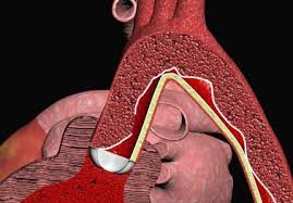
What happens during an aortic dissection?
The aorta carries blood from the heart to the rest of the body. It rises from the heart, curves through the chest and descends toward the abdomen. Its branches supply the brain, arms, spinal cord, kidneys, bowel and legs. The wall has several layers that normally work together to withstand the pressure of circulating blood. A dissection develops when blood enters and separates layers within that wall. [1]
The original passage for blood is called the true lumen. The new passage within the damaged wall is the false lumen. [15]
An aneurysm means an abnormally enlarged section of artery; a rupture means blood escapes through the vessel wall. These problems can coexist, but the words are not interchangeable. Someone can have an aneurysm without a dissection, and a dissection does not have to begin with a previously diagnosed aneurysm. The emergency concerns are bleeding and interruption of blood flow to organs. [1]
Recognize warning signs without waiting for a textbook pattern
Possible symptoms include abrupt, severe chest, upper-back or abdominal pain, fainting, breathlessness, sudden weakness or difficulty speaking. Pain may spread or change location. Symptoms overlap with heart attack, stroke and other emergencies, so a person cannot safely choose between those diagnoses at home. The priority is emergency assessment, even when the person has no known heart or aortic condition. [3]
Descriptions such as tearing or ripping are familiar, but real symptoms vary. People may describe crushing, sharp, aching or otherwise unusual pain. A pain description alone does not establish the diagnosis or make it safe to dismiss. The NHLBI also highlights visual disturbance, faintness, difficulty walking and symptoms resembling a heart attack or stroke. Tell emergency clinicians what happened in your own words rather than trying to match a phrase from a website. [2]
Relevant background includes a known enlarged aorta, previous aortic treatment and relatives with aortic disease. Share that information promptly if it is available, but do not spend time searching old records before calling for help. The absence of a known family condition is not reassurance: a dissection may be the first event that brings an underlying aortic problem to medical attention. [2]
Occasionally, a dissection occurs without the severe pain people expect. An International Registry of Acute Aortic Dissection study documented patients whose presentation included fainting, heart failure or stroke despite no reported pain. This was an observational hospital registry, not a way to calculate an individual person’s risk. Its practical lesson is that missing the classic symptom does not remove the possibility of serious aortic disease. [5]
A person who collapses or develops sudden neurological symptoms needs urgent assessment on those symptoms alone. New speech difficulty, one-sided weakness or sudden vision loss should never be watched at home while waiting to see whether chest pain appears. Temporary improvement cannot establish that the cause is harmless. Emergency teams assess the full pattern; symptom lists cannot exclude dissection. [5]
Stanford type A, type B and related aortic syndromes
The Stanford classification describes which part of the aorta is involved. Type A means the dissection involves the ascending aorta, the portion leaving the heart, regardless of where the first tear began. It may continue through the arch and far down the descending aorta. Type B means the ascending aorta is not involved. The extent of pain cannot identify the type, and a tear beginning farther down can still become type A if it extends upward. [6]
Specialists may use additional labels for dissections centered on the arch, including non-A non-B, and more detailed systems that describe the entry tear and affected branches. Acute, subacute and chronic describe time since onset, not safety. Treatment also depends on organ blood supply, bleeding, aortic dimensions and the person’s condition. [6]
Aortic dissection belongs to the group called acute aortic syndromes. Another member is intramural hematoma, in which blood collects within the aortic wall, sometimes without a clearly visible communicating tear on the initial scan. A penetrating atherosclerotic ulcer occurs when an atherosclerotic lesion extends into deeper layers of the wall. These conditions can progress or coexist, and each needs specialist assessment based on location, symptoms and imaging. [7]
A report saying hematoma or ulcer instead of dissection does not automatically describe a mild problem. Conversely, the name alone cannot tell a family which procedure is necessary. The 2024 ESC guidance considers the whole clinical picture and includes selected preventive intervention for high-risk uncomplicated type B disease. The distinction between an emergency repair and a planned later intervention should therefore be explained explicitly, including what finding would change the plan. [7]
Who is at risk, and what can be learned afterward?
High blood pressure and aging are important risk factors, but aortic dissection also occurs in younger people. A bicuspid aortic valve, meaning a valve with two leaflets rather than the usual three, and narrowing of the aorta are relevant congenital conditions. Certain inherited connective-tissue disorders affect the strength of the aortic wall. Other contexts include significant chest trauma, previous cardiac or aortic procedures and inflammation involving blood vessels. [8]
These associations do not mean every person with a risk factor will have a dissection. They also do not justify dismissing symptoms in someone who seems healthy. During follow-up, clinicians look for a plausible underlying cause and associated conditions rather than assuming there was one avoidable trigger. A careful history should include earlier scans, valve problems, major injuries and operations. The result can change which parts of the circulation need monitoring and whether relatives should be assessed. [8]
The Oxford Vascular Study examined aortic dissection across a defined population, including events outside specialist hospital registries. It found substantial problems with blood-pressure control before dissection, emphasizing why established hypertension deserves attention even when it causes no symptoms. Population studies are valuable because hospital-only reports can miss people who die before reaching treatment. They still cannot tell an individual exactly when or whether a dissection will happen. [9]
This evidence supports treating blood pressure as a long-term health issue rather than a number that matters only during illness. Review persistent elevation with a clinician and address barriers such as side effects, cost or difficulty taking medicines consistently. Many factors interact, and events can occur without an obvious preventable explanation. Risk reduction is not a guarantee. [9]
Genetic assessment may be particularly useful when dissection occurs at a younger age, when characteristic features suggest an inherited disorder, or when relatives have aortic aneurysm, dissection or unexplained early death. Marfan syndrome and Loeys-Dietz syndrome are examples, but inherited thoracic aortic disease can also occur without obvious outward features. A specialist combines clinical findings, a family history and, where appropriate, genetic testing. [10]
A negative genetic test does not always exclude inherited risk, because not every cause is identifiable. A result of uncertain significance is not the same as a confirmed disease-causing variant. Parents, siblings and children may need aortic imaging even when no causative variant has been found. When a pathogenic familial variant is identified, targeted testing can guide relatives’ care. Genetic counseling helps explain the meaning of a result, its limitations and the next steps for that particular family. [10]
Pregnancy needs a specific aortic care plan
People with known aortic disease or a relevant inherited condition should discuss pregnancy before conception whenever possible. Pregnancy and the period after delivery can change aortic risk, and previous repair does not necessarily remove risk in untreated segments. The 2025 ESC pregnancy guidance recommends coordination between an experienced aortic team and a Pregnancy Heart Team. Counseling should consider the specific diagnosis, prior events, current imaging and the person’s wishes. [11]
The plan may include repeated imaging, a review of medicines and decisions about where and how delivery will be managed. Some medicines need reconsideration in pregnancy, so this belongs in a specialist conversation rather than an independent change at home. Follow-up remains important after birth. Sudden severe chest, back or abdominal pain, collapse or stroke symptoms during pregnancy or postpartum still warrant an immediate emergency call, with the pregnancy or recent delivery mentioned to responders. [11]
How clinicians diagnose a suspected dissection
Diagnosis begins with urgent clinical assessment, including the timing and character of symptoms, relevant medical history and examination. Clinicians may compare pulses and blood pressure, check neurological function and look for signs of inadequate organ circulation. None of those bedside findings is present in every case. Similar arm readings, a normal-looking person or improvement in pain cannot independently exclude a dissection. [12]
An electrocardiogram and blood tests may help assess other dangerous causes of symptoms, but they are not substitutes for aortic imaging when suspicion remains. A heart attack can also coexist with a dissection. Professional diagnostic pathways combine multiple findings and are intended for trained clinicians assessing a patient directly. They should not be converted into a home checklist or online score that tells a symptomatic reader to stay away from emergency care. [12]
CT angiography is commonly used urgently because it can show the location and extent of the dissection, branch-vessel involvement and complications. The scan often examines more than the painful area, because the abnormality may extend along the aorta. Contrast helps clinicians see the blood-filled channels. The imaging team weighs speed, image quality, kidney function, allergies and the person’s stability when selecting the test. [13]
Transesophageal echocardiography, which uses an ultrasound probe in the esophagus, can provide important information near the heart and may be valuable when moving an unstable patient is difficult. MRI can also assess the aorta, although access, examination time and monitoring requirements affect its emergency use. A routine chest X-ray or a normal bedside ultrasound does not reliably rule out the condition. The appropriate examination is the one that answers the urgent clinical question safely and promptly. [13]
D-dimer is a blood marker used in some hospital diagnostic pathways. The ADvISED prospective study examined it together with a clinical risk assessment in patients already being evaluated for possible acute aortic syndrome. Combining information helped identify selected patients with a low likelihood of disease, but false-negative cases occurred. An isolated negative blood result is therefore not a universal clearance test for dissection. [14]
A positive result is also nonspecific and may occur for other reasons. The question is whether the test is appropriate within a validated clinical pathway, not whether a person can obtain a reassuring number. When clinical suspicion is high, urgent imaging should not be delayed in the hope that a blood test will settle the issue. Readers should not use a laboratory report, a pulse check or a symptom questionnaire to override an emergency assessment. [14]
Immediate hospital care and type A surgery
Hospital treatment aims to protect the circulation while the aortic team determines the safest repair or management strategy. In suitable patients, closely monitored treatment reduces heart rate, blood pressure and the force acting on the damaged wall. Clinicians also treat pain and watch for changes in consciousness, urine output and circulation. People who are bleeding or in shock require a different resuscitation approach, which is why treatment cannot be reduced to a single blood-pressure target for everyone. [16]
Care may involve transfer to a specialist center with cardiac and vascular surgery, advanced imaging, anesthesia and intensive-care expertise. Transfer arrangements are clinical decisions based on urgency and stability. Families can help by providing a contact number and available information about previous procedures or serious allergies. Some patients go directly to an operating theater or intensive-care unit on arrival. Ask which team will provide updates and where relatives should wait. [16]
Acute type A dissection generally requires emergency open surgery, with immediate surgical assessment. The ascending aorta is close to the heart, aortic valve and coronary arteries. Damage here can cause major bleeding, pressure around the heart, valve leakage or loss of blood supply to the heart and other organs. Medicines are an essential part of stabilization, but they do not replace the recommended emergency repair in an appropriate surgical candidate. [15]
Open repair replaces or reconstructs the damaged section using a surgical graft. The exact operation depends on the involved aorta and whether the valve, root, arch or nearby vessels also need treatment. Endovascular repair takes a different route: a catheter carries a stent graft through an artery, commonly from the groin, to support the affected aortic segment. This is often called thoracic endovascular aortic repair, or TEVAR. [17]
A stent graft can cover an entry tear and improve flow through the true lumen. Some patients need additional treatment to restore blood flow into branch arteries. Endovascular treatment is not suitable for every anatomy and is not the routine substitute for acute ascending-aortic surgery. Potential complications include bleeding, stroke, spinal-cord injury, kidney injury and further aortic problems. Smaller access incisions do not make it a minor procedure or remove the need for continuing surveillance. [17]
Type B treatment: complications determine urgency
An uncomplicated acute type B dissection is usually treated initially with intensive medical management and close observation. Uncomplicated means the team has not identified features such as rupture or threatened organ blood supply; it does not mean the dissection is harmless. Blood-pressure and heart-rate control, symptom reassessment and repeat imaging are active treatment. The situation can change, especially early after the event. [18]
Complicated type B disease may involve rupture, malperfusion that reduces organ or limb blood supply, progression or enlargement, persistent pain, or blood pressure that remains difficult to control despite treatment. Urgent intervention is often required. TEVAR is commonly favored when suitable, while open surgery or other procedures remain necessary in some patients. Underlying connective-tissue disease and the ability to achieve a durable repair can influence the choice. A specialist team should explain which complication is driving its recommendation. [18]
There is an important difference between treating an urgent complication and placing a stent preventively in an otherwise uncomplicated case. The 2026 ESVS guideline does not support routine TEVAR for all uncomplicated acute type B dissections outside clinical trials. It supports continued medical treatment and imaging, while allowing individualized consideration of intervention in selected patients, including those with concerning enlargement. Timing may favor the subacute phase when the situation permits. [19]
Recommendations about preventive repair are more selective than a simple rule that all type B dissections need stents or none ever do. The aortic team weighs imaging features, symptoms, procedural hazards, life expectancy and the strength of available evidence. Useful questions include what benefit is expected now, what would trigger a later procedure, and how the alternative will be monitored. A proposed period of observation should come with a defined clinical and imaging plan. [19]
The INSTEAD-XL report followed 140 patients with stable type B dissection who had previously been randomized to medical treatment with or without TEVAR. Extended follow-up suggested advantages for aorta-related outcomes and disease progression in the intervention group. This was a selected study population and a long-term analysis of a relatively small trial, rather than proof that every newly diagnosed patient should receive an immediate stent. [20]
The timing of enrollment and the outcomes measured matter when interpreting that result. Prevention of later aortic events is a different question from surviving the first hours of a complicated dissection. Treatment also has immediate risks. Ask how closely your anatomy and clinical situation match the trial evidence. [20]
The ADSORB trial addressed a related question in uncomplicated acute type B dissection. It randomized 61 patients to medical care alone or medical care plus a stent graft. At one year, the stent group showed more favorable changes in aortic structure, including the false lumen. This process is called remodeling: it describes how the channels and aortic dimensions change over time. [21]
A favorable scan result is encouraging, but it is not identical to a demonstrated improvement in survival or daily functioning. This small trial was not large enough to establish a definitive mortality advantage. Together, these studies explain why specialists discuss both potential long-term benefit and procedural risk, and why further research remains useful. Trial headlines should not override an individualized clinical plan. [21]
Understanding prognosis without a misleading countdown
Aortic dissection can deteriorate suddenly, so rapid assessment and treatment matter. However, a single hourly death rate cannot describe every dissection. A contemporary IRAD analysis of acute type A disease found that early outcomes differed substantially between patients intended for surgery and those managed medically. Those groups also differed in age and illness, making simple comparisons difficult. The study included patients recognized at participating hospitals, which affects how broadly its results apply. [22]
For an individual, prognosis depends on the involved aorta, rupture, shock, organ injury, other illnesses and whether effective treatment can be delivered. An initially stable patient may still worsen unexpectedly. Equally, frightening population statistics should not be treated as a prediction that a particular person cannot survive or recover. Ask the treating team about the current organ findings, the purpose of the proposed treatment and the main uncertainties. Emergency urgency is clear without inventing a universal countdown. [22]
Lifelong imaging, medicines and repeat treatment
Surviving the emergency or receiving a graft does not necessarily remove disease from the rest of the aorta. Residual dissection can remain, and affected segments may enlarge over time. The American Heart Association’s statement on chronic dissection emphasizes lifelong surveillance. Imaging looks at the remaining aorta and relevant branches, the repair and changes that might justify additional treatment before another emergency occurs. [23]
Accurate comparison requires more than reading two diameter numbers from separate reports. Measurements can vary with scan technique, the point measured and image orientation. Specialists may need the actual previous images to decide whether growth is real. Keep a record of the imaging facility and dates, and ask how records will be transferred if care changes. CT and MRI each have advantages; the surveillance plan balances the information needed with contrast, radiation, access and individual medical circumstances. [23]
For dissection managed medically, and after repair when residual aortic disease remains, the 2022 ACC/AHA guideline recommends CT or MRI at one month, six months and twelve months, then annually if stable. Individual circumstances can require earlier or more frequent studies. Confirm your own schedule before discharge; a general timetable cannot replace the treating team’s instructions. [15]
A population-based Ontario study followed survivors after surgery for acute type A dissection and documented later aortic procedures over years of follow-up. Some required urgent rather than planned treatment. This does not mean every repair eventually fails. It shows why the story does not end when the chest wound heals and why a future procedure can be part of managing a continuing aortic condition. [24]
The study also examined how consistently surveillance imaging occurred. Observational findings cannot establish a perfect scan schedule or prove that one missed scan causes a later complication. For patients, the practical step is to identify who owns follow-up and to contact that team when an expected appointment or scan is missing. Arrange an explicit handover if hospitals or clinicians change. [24]
Long-term medicines commonly help reduce stress on the aorta by controlling blood pressure and, where appropriate, heart rate. A Taiwanese observational study involving nearly seven thousand dissection survivors found associations between some antihypertensive drug groups and better later outcomes. Because treatment was not randomly assigned, differences between patients could partly explain those findings. The study does not identify one universally best prescription for every survivor. [25]
The clinical plan depends on the repair, residual disease, kidney function, other conditions and medication tolerance. Ask for a clear list explaining what each medicine is for and who will review it. Report dizziness, excessive fatigue, affordability problems or difficulty following the regimen rather than silently stopping treatment. Do not change doses using an article or another patient’s experience. Prescription review is part of continuing care, especially when another clinician adds a medicine. [25]
If home blood-pressure monitoring is recommended, ask the team which arm to use and what readings require contact. A validated automatic upper-arm monitor and correctly fitting cuff improve reliability. For routine measurements, rest quietly first, support the back and arm, keep feet on the floor and place the cuff on bare skin at heart level. Avoid talking during the reading. Follow the team’s advice about timing and frequency. [26]
The American Heart Association recommends taking two readings one minute apart and recording them. Bring the monitor and record to appointments so technique and results can be reviewed. These measurements help manage a known condition; they cannot diagnose or exclude a new dissection. If emergency symptoms occur, call immediately rather than repeatedly checking the cuff. A normal number does not make sudden severe pain, collapse or stroke symptoms safe to monitor at home. [26]
Recovery, movement and emotional health
Recovery depends on the emergency itself, the procedure, organ injury and previous health. After open surgery, wound and breastbone healing add demands that may differ from recovery after an endovascular procedure or medical treatment. Before discharge, obtain instructions for wound care, activity, medicines, follow-up and the symptoms that need urgent contact. Ask who is available outside routine clinic hours and make sure someone helping at home understands the plan. [30]
Discuss work, driving, travel and caring responsibilities with the team rather than using another patient’s timeline. A desk job and heavy physical work pose different demands. Driving also involves safe control of the vehicle and applicable local rules. Arrange practical help with meals, transport or household tasks. Bring questions about sleep, stamina and daily activities to follow-up. [30]
Activity advice should be individualized. A small study of adults with thoracic aortic aneurysms or dissections measured blood-pressure responses during a supervised moderate-exercise protocol and found that this level of activity was feasible for many participants. It did not establish a universal safe lifting limit or prove that strenuous exercise is safe for every person with a damaged aorta. The findings support assessment and guidance rather than either blanket inactivity or unrestricted training. [27]
Ask about the activities you actually want to do: walking, cycling, gardening, carrying groceries or returning to a sport. The answer may depend on the remaining aorta, blood-pressure control and recovery stage. Heavy straining, breath-holding and sudden maximal effort deserve specific discussion. A supervised plan can establish appropriate intensity and progression, including what symptoms mean stopping and seeking help. Do not copy a research exercise protocol as a home test of whether your aorta can tolerate exertion. [27]
Cardiac rehabilitation can provide a structured bridge between hospital treatment and everyday activity. A small retrospective pilot study after type A surgery found improvements in exercise capacity and reported quality of life among participants who completed an exercise-based program. The study was encouraging but limited by its size and nonrandomized grouping. It cannot promise the same improvement for every survivor or determine the ideal start date for every procedure. [28]
Ask whether a rehabilitation service familiar with aortic disease is available and whether your team recommends referral. The program should know about residual dissection and any restrictions, rather than treating the operation as an ordinary exercise setback. If travel, cost or scheduling makes attendance difficult, raise that early so alternatives can be considered. Goals can include confidence with daily movement and a sustainable routine, with progress adjusted to symptoms and medical review. [28]
Emotional recovery can take longer than relatives expect. Research using questionnaires after emergency type A surgery has documented distress, reduced well-being and symptoms suggesting post-traumatic stress in some survivors. A questionnaire result is not itself a psychiatric diagnosis, and survey respondents may differ from people who did not respond. Still, the evidence supports taking these experiences seriously rather than assuming that a successful operation ends the psychological effects. [29]
Tell the care team about persistent fear, intrusive memories, disturbed sleep or avoidance that interferes with daily life. A clear explanation of the follow-up plan may help, and some people benefit from psychological treatment or peer support. Families may also need help understanding what happened and how to support recovery without creating constant alarm. New physical warning symptoms still require medical assessment; they should not automatically be attributed to anxiety because someone has survived a frightening event. [29]
Build a prevention and follow-up routine you can sustain
Prevention focuses on modifiable risks and known aortic disease. Smoking cessation, appropriate blood-pressure management and attention to other cardiovascular conditions are useful parts of care. An eating pattern that supports cardiovascular health can help with those goals, but no food, supplement or cleanse can seal a dissection. Physical activity advice must be adapted to the aortic diagnosis and recovery stage rather than copied from general fitness recommendations. [31]
If an aneurysm was found before a dissection, scheduled monitoring and appropriately timed treatment are intended to reduce future complications. The decision depends on more than one diameter measurement: the location, growth pattern, underlying cause and overall health matter. Stay connected with the clinician responsible for that assessment. Prevention is most workable when the plan also addresses everyday barriers, including access to appointments and help with smoking cessation, rather than relying on willpower or a single reassuring scan. [31]
Frequently asked questions
What should I do about possible aortic dissection symptoms?
Call your local emergency number immediately. Do not drive yourself.
Does aortic dissection always cause tearing pain?
No. Pain can vary, and some dissections are painless.
What defines Stanford type A?
The dissection involves the ascending aorta.
Does type B always need immediate surgery?
No. Specialists assess complications and anatomy.
Is follow-up needed after successful aortic repair?
Yes. Lifelong aortic surveillance remains necessary.
Should close relatives discuss aortic screening?
Ask about family imaging and genetic assessment.
Sources
This educational guide was checked against the sources below on 8 October 2026. It does not replace emergency assessment or an individual care plan. No clinician certification is claimed. Read our medical disclaimer.
- AHA: Understanding the aorta and aortic emergencies
- NHLBI: Aortic dissections, risks and recognition (2024)
- Mayo Clinic: Aortic dissection symptoms and causes
- NHS: Fainting and emergency action
- Park and colleagues: Painless acute aortic dissection (2004)
- EACTS/STS: Aortic organ guidelines (2024)
- ESC: Peripheral arterial and aortic disease guidelines (2024)
- MedlinePlus: Aortic dissection
- Howard and colleagues: Oxford Vascular Study (2013)
- GeneReviews: Heritable thoracic aortic disease overview
- ESC: Cardiovascular disease and pregnancy guidelines (2025)
- Ohle and colleagues: Canadian diagnostic guideline (2020)
- ACR: Suspected acute aortic syndrome imaging criteria (2021)
- Nazerian and colleagues: ADvISED diagnostic study (2018)
- ACC/AHA: Aortic disease guideline (2022)
- NHS England: Acute aortic dissection transfer information (2024)
- Society for Vascular Surgery: Dissection treatment information
- STS/AATS: Type B dissection guideline (2022)
- ESVS: Descending thoracic and thoraco-abdominal aortic guidelines (2026)
- Nienaber and colleagues: INSTEAD-XL follow-up (2013)
- Brunkwall and colleagues: ADSORB trial (2014)
- Harris and colleagues: Early type A mortality in IRAD (2022)
- AHA: Imaging and surveillance of chronic dissection (2022)
- JACC: Surveillance after acute type A repair (2021)
- Chen and colleagues: Long-term blood pressure medicines (2021)
- AHA: Home blood pressure monitoring
- Li and colleagues: Exercise in thoracic aortic disease (2022)
- Fuglsang and colleagues: Rehabilitation after type A surgery (2017)
- Study: Quality of life after acute type A surgery (2018)
- Barts Health NHS Trust: Advice when having aortic surgery
- NHLBI: Aortic aneurysm treatment and prevention measures
- HSE: Aortic dissection overview (reviewed 2026)