

Kidney and urinary health
Kidney stones form when substances dissolved in urine build up into crystals that grow and collect together. Their causes include concentrated urine, changes in urine chemistry, certain infections, inherited conditions, and other medical problems. Understanding the stone type helps a clinician explain why it formed and choose treatment and prevention that fit the person.
A stone may remain in a kidney without noticeable symptoms, pass through the urinary tract, or become stuck in a ureter, the narrow tube carrying urine from a kidney to the bladder. A blockage can cause intense pain and sometimes threaten kidney function. Stone size matters, but location, infection, kidney health, and the person’s overall condition also influence how urgently care is needed. [1, 2]
This guide provides general education, not a diagnosis or an individual treatment plan. A healthcare professional should assess new symptoms and tailor advice, especially during pregnancy, in children, or when kidney or heart disease limits fluid intake.
How do kidney stones form?
The kidneys filter the blood and produce urine containing water, minerals, and waste products. These substances normally leave the body without forming stones. Problems can develop when urine contains more of a stone-forming substance than can remain dissolved. Low urine volume makes the same amount of material more concentrated, increasing the opportunity for crystals to develop. A stone often reflects several interacting factors rather than one food or one missed drink.
Urine chemistry matters as well as its volume. Calcium, oxalate, phosphate, uric acid, and cystine have different chemical properties. The acidity or alkalinity of urine, measured as its pH, affects which substances are likely to crystallize. Urine also contains substances such as citrate that help discourage certain crystals from becoming stones. A prevention plan may therefore need to address urine volume, the amount of a particular mineral, and the balance of protective substances. [1, 4, 5]
Kidney stones are sometimes called renal calculi or nephrolithiasis. The broader term urolithiasis describes stones in the urinary tract. These names do not identify the stone’s chemical composition. A stone’s appearance or the intensity of pain cannot reliably tell someone what it contains. Laboratory analysis of a retrieved stone provides more useful information for prevention than judging its color, shape, or a photograph.
A cause is the biological process responsible for a stone, such as excess urinary cystine from an inherited disorder. A risk factor increases the chance of that process producing a stone, such as persistently low urine volume. Someone can have several risk factors and never develop a stone, while another person develops stones despite careful habits. Keeping that distinction clear avoids blame and helps focus testing on explanations that can change care. [1, 3, 10]
For a general biology refresher, explore the Excretion and Homeostasis topics in our biology flashcards.
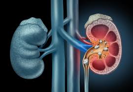
The main kidney stone types and their causes
Calcium oxalate stones
Calcium stones are the most common category, and calcium oxalate is the most common form within that category. They can develop when urine contains too much calcium or oxalate, too little citrate, or insufficient water to keep these substances diluted. Some people excrete extra calcium in urine because of an inherited tendency or an underlying condition. This does not automatically mean that their blood calcium is high or that they eat excessive calcium. [1, 5]
Oxalate comes from some foods and from the body’s own metabolism. Digestive conditions can increase how much is absorbed. In rare inherited disorders called primary hyperoxaluria, the body produces excessive oxalate, creating a different problem from ordinary dietary intake. Identifying the mechanism matters: a person with intestinal disease, someone with high urine calcium, and someone with primary hyperoxaluria may all form calcium oxalate stones but need different investigations and care. [1, 13]
Calcium phosphate stones
Calcium phosphate stones also contain calcium, but their formation is particularly influenced by alkaline urine. Some occur with renal tubular acidosis, a disorder affecting the kidneys’ handling of acid, or hyperparathyroidism, which can disturb calcium balance. Certain calcium phosphate minerals can be associated with infection, while others are not. The laboratory report and blood and urine results help distinguish these situations. A plan designed for calcium oxalate stones should not simply be copied without considering these differences. [8, 11, 12]
Uric acid stones
Uric acid is a waste product formed when the body breaks down purines, substances found in body tissues and many foods. Uric acid stones form especially readily in persistently acidic urine. A low urine volume or increased uric acid in urine can add to the risk. Gout, metabolic conditions, and a diet containing excessive amounts of certain animal proteins can be relevant, but a person does not need every one of these factors to develop a stone. [1, 5]
The distinction matters because selected uric acid stones can sometimes be dissolved through prescribed treatment that changes urine chemistry. This requires a confirmed or sufficiently supported diagnosis, a safe monitoring plan, and follow-up imaging. It is not a reason to start alkalinizing products independently or assume that a painful obstruction can be managed with drinks alone. Calcium-containing stones generally do not respond to that same dissolution approach. [7]
Struvite and infection-related stones
Struvite stones are linked to certain urinary infections. Bacteria that produce the enzyme urease change the chemical environment of urine, allowing infection-related minerals to form. These stones can enlarge rapidly and may occupy a substantial part of the kidney’s collecting system. A person may need both treatment of the infection and treatment to remove stone material; changing diet by itself does not address the underlying problem. [1, 8]
Not every urinary tract infection produces a struvite stone, and not every stone found during an infection is made of struvite. An ordinary stone can also obstruct urine flow and become associated with a dangerous infection. Clinicians therefore assess infection and obstruction separately from the question of stone composition. A previous label of “calcium stone” does not make fever during a new stone episode safe to ignore. [7, 14]
Cystine stones
Cystine stones result from cystinuria, an inherited condition in which the kidneys do not properly reabsorb certain amino acids. Cystine accumulates in urine and can crystallize. This is a specific genetic cause, not something a person acquires by eating one particular food. Stones may recur, so specialist follow-up often combines a carefully planned fluid strategy with changes in urine chemistry and, when needed, additional medicines. Family assessment may be discussed when the diagnosis is established. [8, 10]
| Stone type | Key contributor | Prevention focus |
|---|---|---|
| Calcium oxalate | Urine calcium, oxalate, citrate, and concentration | Match food and medicine advice to urine findings |
| Calcium phosphate | Urine pH and calcium; sometimes an underlying disorder | Investigate the cause and monitor urine chemistry |
| Uric acid | Especially acidic urine | Clinician-directed urine chemistry and dietary care |
| Struvite | Certain urinary infections | Infection control and appropriate stone treatment |
| Cystine | Inherited cystinuria | Specialist prevention and ongoing monitoring |
Risk factors: why some people develop stones
Low urine volume, heat, and everyday routines
Drinking too little for the body’s needs can leave urine concentrated. Sweat losses during hot weather, physical work, or exercise can contribute even when someone drinks their usual amount. Repeated vomiting or diarrhea can also reduce the water available for urine production. These situations explain why prevention advice must consider a person’s work, climate, activity, and health rather than assume that one fixed number of glasses suits everyone. [4]
Practical barriers deserve attention. A person may avoid drinking because toilets are difficult to access, urinary urgency is troublesome, or work breaks are limited. Discussing those barriers can make a plan more realistic. Someone who has been instructed to restrict fluids because of heart or kidney disease should keep following that instruction and ask the responsible clinician to coordinate stone prevention. Increasing fluid intake without that discussion can be unsafe. [4, 17]
Food patterns, supplements, and medicines
A high sodium intake can increase the amount of calcium passed in urine. Large amounts of animal protein may affect uric acid and other aspects of urine chemistry. High oxalate intake is particularly relevant when someone forms calcium oxalate stones and has increased urine oxalate. The useful question is how the overall eating pattern interacts with the person’s laboratory findings, not whether an isolated food is universally “good” or “bad.” [5]
Medicines and supplements can also matter. Some drugs alter urine chemistry, and others can contribute material that forms stones. Topiramate is one example a clinician may review when assessing stone risk. Calcium-containing antacids and vitamin supplements should also be included in a complete medication history. Do not stop a prescribed medicine because it appears on a risk-factor list; the prescriber can weigh its benefits, alternatives, and any monitoring or prevention needed. [1, 8]
Digestive, hormonal, metabolic, and inherited conditions
Inflammatory bowel disease, chronic digestive problems, and some gastrointestinal operations are associated with stones. In certain conditions, changes in absorption increase the oxalate reaching the kidneys. Ongoing diarrhea can compound the problem through fluid loss. A diet that is suitable for uncomplicated stones may need adjustment when someone also has malabsorption, nutritional deficiencies, or restrictions related to bowel disease. This is a useful setting for coordination between the treating clinician and a registered dietitian. [1, 5]
Hyperparathyroidism can raise blood calcium and increase stone risk. Renal tubular acidosis can disturb acid balance and contribute to kidney stones and other complications. Gout, obesity, and metabolic conditions can also be relevant to the assessment. Finding one of these conditions changes the conversation from general prevention to treating a specific medical contributor. It does not establish the diagnosis from symptoms alone; appropriate blood tests, urine testing, and clinical interpretation are needed. [1, 11, 12]
Previous stones and a family history increase the likelihood of future stones. Family history can reflect shared habits, inherited differences in mineral handling, or a less common genetic disorder. Early onset, repeated episodes, or a known inherited condition are reasons to discuss whether more detailed evaluation is useful. A relative’s experience can provide helpful history, but their stone type, medication, and diet should not be assumed to apply to someone else. [1, 10, 13]
Symptoms and warning signs
The characteristic symptom is pain in the side or back that may spread toward the lower abdomen or groin. It can fluctuate in waves and be accompanied by nausea or vomiting. Blood in the urine may be visible as pink, red, or brown discoloration, or detectable only by a test. Needing to urinate frequently, pain during urination, and passing only a small amount of urine can also occur. Small stones sometimes cause no symptoms. [2]
These symptoms overlap with urinary infections and other abdominal or pelvic conditions. Even someone who has had a stone before should not assume that every episode of similar pain has the same explanation. A new episode needs assessment appropriate to its severity, associated symptoms, and the person’s medical history. The absence of visible blood does not provide enough reassurance to dismiss severe pain or other concerning changes.
Fever or shaking chills with possible stone symptoms raises concern about infection. If infection occurs behind an obstruction, bacteria and infected urine may not drain normally, and the illness can progress to sepsis. Confusion, difficulty breathing, or feeling acutely and seriously unwell warrants emergency help. Inability to pass urine also needs emergency assessment. A stone does not have to be large to create a clinically important blockage. [2, 7, 16]
Uncontrolled pain, repeated vomiting, or inability to keep fluids down needs urgent care rather than prolonged self-treatment. Tell the service immediately about pregnancy, a single functioning kidney, significant kidney disease, or immune suppression, because these details can affect urgency and treatment. If a clinician has already diagnosed a stone, follow the specific return precautions provided; worsening symptoms require reassessment even when a follow-up appointment is already booked. [4, 6, 7]
How kidney stones are diagnosed
History, examination, and laboratory tests
Assessment starts with the pattern of pain, urinary symptoms, fever, vomiting, and previous stone episodes. Clinicians also ask about medicines, supplements, relevant illnesses, surgery, family history, and pregnancy possibility when applicable. Examination and vital signs help assess how unwell someone is and whether another diagnosis needs consideration. The immediate priority is identifying dangerous complications; the longer-term investigation asks why stones are forming. [3]
A urine test can look for blood, crystals, and signs of infection. A urine culture may be needed to identify bacteria and guide antibiotic selection. Blood tests can assess kidney function and abnormalities in minerals such as calcium, alongside other tests indicated by the clinical picture. Results are interpreted together: a single abnormal value does not always establish a cause, and treatment of an urgent problem should not wait for every prevention test to be completed. [3, 7]
Imaging: why the choice varies
Imaging establishes whether a stone is present, where it is, how large it is, and whether urine flow is obstructed. Non-contrast CT provides detailed information and is commonly used for adults with suspected renal colic. It involves radiation, so clinicians consider whether CT is needed and whether a lower-dose technique is appropriate. Ultrasound avoids radiation but can leave uncertainty that requires further investigation. Local pathways and the patient’s circumstances influence the first test. [3, 6]
Ultrasound is the preferred initial examination in pregnancy and for children. During pregnancy, a specialist team may consider MRI when ultrasound is inconclusive; low-dose CT is reserved for selected situations when needed after weighing the benefits and risks. A plain abdominal X-ray is useful for some stones and follow-up questions, but not every stone is visible on it. An imaging report should therefore be interpreted in the context of the test used. [3, 6, 7]
Stone analysis and a prevention assessment
If a stone is passed or removed, laboratory analysis can identify its composition. A clinician may provide a urine strainer and instructions for collecting and storing it. Retrieving a sample can avoid guesswork, but nobody should postpone necessary care while trying to capture a stone. Keep the analysis result with other medical records, especially when changing clinicians or seeking care during travel. [4]
After the acute episode, some people are asked to collect urine over a full day to measure volume and stone-related chemicals. This is different from the small urine sample used to check an infection. The collection can help identify a preventable pattern and assess whether treatment is working. Ask exactly when to collect, how to handle a missed sample, and whether to follow the usual routine. An incomplete collection can make results harder to interpret. [4, 8]
Blood and urine measurements answer different questions. A blood result helps describe what is circulating in the body, while a urine collection shows what the kidneys are excreting over the collection period. Normal blood calcium therefore does not, by itself, settle whether urine calcium contributes to stones. Likewise, identifying a calcium-containing stone does not tell the clinician which of several possible contributors is present. Ask the team to explain how the results fit together and which findings would actually change the prevention plan. [1, 3, 8]
Treatment depends on the stone and the person
Observation, symptom relief, and assisted passage
Some small stones pass without a procedure. Observation is an active plan that includes symptom treatment, clear return precautions, and appropriate follow-up. The likelihood of passage depends on location and size, while the safety of waiting depends on infection, kidney function, ongoing obstruction, and whether symptoms can be controlled. A person who initially qualifies for observation may need a different plan if the clinical situation changes. [4, 7]
Pain relief and treatment for nausea are selected according to medical history and the immediate situation. An over-the-counter medicine that is suitable for one person may be unsuitable for someone with kidney disease, stomach bleeding risk, pregnancy, or interacting medicines. Follow the clinician’s directions rather than combining products or increasing doses independently. People who are vomiting or dehydrated may need hospital treatment, including fluids when clinically appropriate. [4, 6]
A clinician may offer a medicine to help selected ureteric stones pass. Suitability depends on the stone and the individual’s risks; this does not mean every kidney stone benefits from the same drug. Ask what benefit is expected, which side effects matter, and when the plan will be reviewed. Do not attempt to force passage by drinking excessive water quickly, particularly if urine output is low or vomiting is persistent. Evidence does not establish a reliable benefit from forced high-volume fluids during acute ureteric colic. [6, 7, 18]
Procedures to break up or remove stones
Shock wave lithotripsy uses energy delivered from outside the body to break a stone into smaller pieces that can pass through the urinary tract. Ureteroscopy uses a thin instrument passed through the urinary opening and bladder into the ureter or kidney, allowing a stone to be removed or fragmented. Both approaches have specific uses, and the choice depends on more than the stone’s diameter. [4]
Percutaneous nephrolithotomy reaches the kidney through a small opening in the back and is an option for larger or more complex stones. The urologist explains expected benefits, anesthesia, recovery, and potential complications of the proposed procedure. Ask whether more than one session may be needed and how the team will check for remaining fragments. Removing the present stone addresses that episode; prevention may still be needed afterward. [4]
A temporary ureteral stent may be left to help urine drain. It can produce urinary symptoms, so the discharge plan should explain expected effects and which changes need attention. Confirm who will remove or exchange it and when. A stent is a medical device with a follow-up plan, not something to forget once pain improves. If appointment details are missing, contact the treating service for clarification. [4, 19]
Why infection with obstruction changes the plan
An infected, obstructed kidney is a urological emergency. Treatment can require urgent drainage using a ureteral stent or a tube placed directly into the kidney, together with antibiotics and other hospital care. Definitive stone removal is commonly arranged after infection is controlled. Antibiotics alone may not solve the problem when infected urine cannot drain. This is why fever during a stone episode requires urgent assessment rather than waiting for routine treatment. [7]
Preventing future stones without a one-size-fits-all diet
Agree on a safe fluid plan
For many people, producing enough urine to dilute stone-forming substances is a central prevention goal. Water is usually the simplest main drink. The amount needed varies with body size, climate, activity, losses from illness, and other health conditions. A clinician may use a timed urine collection to assess whether the plan is producing enough urine instead of relying only on an estimated daily drink total. [4, 5]
Spread an agreed fluid intake across daily routines in a way that is practical and sustainable. Access to water at work or school, a refillable bottle, and a plan for hot days can help. Pale urine can be a rough clue about concentration, but urine color is affected by other factors and does not prove that stone risk is controlled. Follow any prescribed fluid restriction and ask the clinician managing heart or kidney disease to help set the target. [4, 17]
Citrus drinks may supply citrate, but their composition varies, and sugar or other ingredients can matter. Lemon water is not a reliable substitute for prescribed citrate medicine or a way to treat an infected obstruction. There is also no reason to start drinking alcohol to prevent stones. Choose drinks within an overall healthy eating plan and discuss special requirements with the care team. [4, 5]
Keep appropriate dietary calcium and review sodium
A calcium-containing stone does not automatically call for a low-calcium diet. Calcium in food can bind oxalate in the digestive tract, reducing how much oxalate is absorbed. Unnecessary restriction can also conflict with bone health and, in children, growth. The appropriate intake depends on age and other conditions. A clinician or dietitian can help select suitable foods without treating every dairy product or calcium-containing food as a cause of stones. [5, 14]
Calcium supplements require a separate discussion from ordinary food intake. Their purpose, amount, and timing may matter, particularly in people with digestive disorders or nutritional needs. Bring all supplements to the review rather than assuming “natural” products are irrelevant. Sodium reduction can be useful for calcium stone prevention; comparing food labels and noticing sodium in packaged foods, sauces, and restaurant meals may be more informative than focusing only on salt added at the table. [1, 5]
Match oxalate and protein advice to the findings
People with calcium oxalate stones and high urinary oxalate may need to adjust foods that supply substantial oxalate, such as spinach, rhubarb, or certain nuts and nut products. The amount and frequency eaten matter. A restrictive internet list that removes many nutritious foods may be unnecessary or difficult to sustain. Dietitian support is especially helpful when other medical conditions already limit food choices. [5]
Animal protein advice should also be individualized. Excessive intake may contribute to stone risk, particularly for uric acid stones, but everyone still needs adequate nutrition. Replacing some portions with suitable plant proteins may help some people. The choice should account for stone type, other illnesses, and nutritional requirements. Avoid adopting a highly restrictive or high-protein weight-loss diet without discussing its implications when there is a history of stones. [5]
Use stone-specific medicines and address underlying disease
Prevention sometimes requires medication as well as food and fluid changes. Clinicians may consider treatments that reduce urinary calcium, increase citrate, change urine pH, or reduce uric acid, depending on the findings. These medicines have different purposes and potential adverse effects. Kidney function, other prescriptions, and follow-up blood or urine tests help determine which option is suitable. A medication used by a friend with stones is not a safe shortcut to a personal prevention plan. [4]
Calcium phosphate stones need particular attention to the cause and urine chemistry. Uric acid stones often require attention to urine acidity. Infection-related stones require infection control and appropriate stone treatment, while cystinuria needs ongoing specialist prevention. Renal tubular acidosis, hyperparathyroidism, and primary hyperoxaluria each require their own assessment and treatment. These differences explain why “drink more water” can be useful general advice yet remain an incomplete plan for an individual. [8, 10, 11, 12, 13]
Children, pregnancy, and family considerations
Children can develop stones, including those without a recognizable adult pattern of risk factors. Symptoms can include abdominal or side pain, blood in urine, vomiting, or irritability in younger children. A child with these symptoms needs medical assessment; parents should not apply an adult treatment or fluid target. Evaluation considers growth, nutrition, urinary anatomy, family history, and possible metabolic or inherited conditions. A pediatric team can arrange prevention that remains appropriate as the child grows. [14, 15]
School arrangements may be part of making that plan workable. Families can ask about access to drinking water, permission to use the toilet, and instructions for responding to pain or illness. The clinical team should guide any dietary changes so that prevention does not create unnecessary food anxiety or nutritional gaps. If urine collection is requested, parents can ask for age-appropriate instructions and help with practical difficulties. [14]
During pregnancy, new flank or abdominal pain should be assessed rather than assumed to be a stone. Imaging and treatment choices must account for both the pregnant person and the pregnancy. Ultrasound is usually the first imaging test, and the obstetric and urology teams coordinate when symptoms persist or complications occur. Do not use medicines left over from an earlier stone episode without checking whether they are suitable in pregnancy. [6, 7]
A family history is useful information rather than a prediction of inevitable stones. Tell the clinician about relatives with childhood-onset stones, repeated procedures, or a known diagnosis such as cystinuria or primary hyperoxaluria. Genetic evaluation may be appropriate in selected situations. The decision depends on the pattern of illness and test results; sharing a surname or having one affected relative is not enough to diagnose an inherited disorder. [1, 10, 13]
For general biology learning about the molecules involved in inheritance, see our nucleic acids study guide.
Follow-up, recurrence, and protecting kidney health
People who have had a kidney stone are more likely to develop another. Recurrence risk varies, so a universal percentage cannot describe every person’s outlook. Stone composition, previous episodes, remaining fragments, urinary abnormalities, and underlying illness all matter. Prevention aims to reduce the chance of future stones and complications; it cannot guarantee that another stone will never form. A recurrence should prompt review of the plan, not an assumption that someone failed to follow instructions. [1, 9]
Improvement in pain alone does not always establish that the stone has passed or that obstruction has resolved. Ask how the treating team will confirm the outcome and whether a follow-up scan is needed. Imaging choices balance the clinical question with radiation exposure. Some people can be followed with ultrasound or an X-ray suitable for their stone type, while others need CT for particular symptoms or treatment planning. [9]
Follow-up also provides a chance to review stone analysis, blood results, urine collections, medicines, and any obstacles to the prevention plan. Bring a record of new symptoms and relevant changes, such as gastrointestinal surgery, a new supplement, or a different work environment. If a medicine was started, ask what monitoring it requires and whom to contact about side effects. The timing of review should reflect the person’s risk rather than an arbitrary schedule. [4, 9]
Most people do not develop permanent kidney damage when stones are recognized and appropriately treated, but persistent obstruction, infection, and some underlying disorders can harm kidney function. Keeping follow-up appointments is especially important when symptoms are mild or absent but a stone remains. A clear plan should identify who is responsible for reviewing results, arranging imaging, and managing any temporary stent or drainage tube. [1, 4, 9]
Useful questions for a healthcare appointment
Ask what is known about the stone’s size, location, composition, and effect on urine flow. Clarify whether infection or reduced kidney function changes the urgency of treatment. If observation is proposed, discuss what symptoms should trigger urgent reassessment and how passage will be confirmed. If a procedure is recommended, ask why it fits the current stone and what follow-up will be required.
For prevention, ask whether stone analysis or a timed urine collection would change the plan, what fluid intake is safe alongside other conditions, and which dietary changes have a clear purpose. Discuss whether medications or supplements could contribute, without stopping them independently. Before leaving, confirm how results will be communicated and who to contact if symptoms return. These practical details turn general advice into a plan that can be followed.
Frequently asked questions
1. What is the main cause of kidney stones?
Stones form when urine conditions allow certain substances to crystallize. Concentrated urine is a common contributor, but stone type, urine chemistry, infections, inherited disorders, and other medical conditions matter. Testing may identify several contributors rather than one cause. [1, 2]
2. Should everyone with calcium stones avoid calcium?
No. Appropriate calcium from food is usually part of prevention and supports bone health. Calcium can bind oxalate in the digestive tract. Ask a clinician or dietitian about the right intake and review supplements separately rather than cutting out calcium on your own. [5]
3. Can drinking water flush out a kidney stone?
Some small stones pass with a clinician-directed observation plan, and adequate hydration can support prevention. Excessive drinking does not guarantee passage or safely treat a blockage. Follow fluid restrictions, and seek urgent care for fever, inability to urinate, uncontrolled pain, or repeated vomiting. [2, 4, 7, 18]
4. How long does a kidney stone take to pass?
Timing varies with size, location, and the person’s circumstances. Some stones pass over days or weeks; others need a procedure. The treating clinician should explain how long observation is appropriate, when to return sooner, and how passage or ongoing obstruction will be checked. [6, 7, 9]
5. Is fever normal when passing a kidney stone?
Fever or shaking chills with possible stone symptoms requires urgent medical assessment. Infection behind a blockage can become a medical emergency and may need antibiotics plus urgent drainage. Do not wait at home for the stone to pass or rely on fever medicine alone. [2, 7]
6. Can kidney stones come back after removal?
Yes. A procedure treats existing stones but may not correct the conditions that caused them. Stone analysis, selected blood and urine tests, follow-up imaging, and individualized food, fluid, or medication advice can help reduce future risk. Prevention lowers risk but cannot guarantee no recurrence. [1, 4, 9]
Sources
Sources checked October 6, 2026. Patient information and clinical guidelines serve different purposes; care decisions require clinical assessment. Source numbering corresponds to the non-clickable markers in this guide.
- NIDDK: Definition & Facts for Kidney Stones
- NIDDK: Symptoms & Causes of Kidney Stones
- NIDDK: Diagnosis of Kidney Stones
- NIDDK: Treatment for Kidney Stones
- NIDDK: Eating, Diet, & Nutrition for Kidney Stones
- NHS: Kidney Stones
- European Association of Urology: Urolithiasis Guidelines, 2026, diagnosis and treatment
- European Association of Urology: Metabolic Evaluation and Recurrence Prevention, 2026
- European Association of Urology: Follow-up of Urinary Stones, 2026
- MedlinePlus Genetics: Cystinuria
- NIDDK: Renal Tubular Acidosis
- NIDDK: Primary Hyperparathyroidism
- MedlinePlus Genetics: Primary Hyperoxaluria
- NIDDK: Treatment & Prevention for Kidney Stones in Children
- NIDDK: Symptoms & Causes of Kidney Stones in Children
- NHS: Sepsis
- North Tees and Hartlepool NHS Foundation Trust: Kidney Stones
- Cochrane: Fluids and Diuretics for Treating Acute Ureteric Colic
- University Hospitals Plymouth NHS Trust: Managing a Ureteric Stent after Ureteroscopy
About the accompanying video: The older TED-Ed animation offers a general introduction. Its discussion of fluids and citrate needs qualification: drinking large volumes quickly does not reliably force an obstructing stone to pass, and lemon or citrate-containing drinks do not dissolve most calcium stones. Selected uric acid stones may dissolve with prescribed alkalinization, monitoring, and follow-up. Fever with stone symptoms or inability to urinate needs urgent medical assessment. [2, 7, 18]