

Fuchs dystrophy affects cells that help keep the cornea clear. Its course varies, and treatment depends on how the eyes are affected. This guide explains the condition, assessment and care options; it cannot diagnose an individual eye problem. [3]
What Fuchs dystrophy changes inside the cornea
The cornea is the transparent, curved window at the front of the eye. Light passes through it before travelling through the pupil and lens toward the retina. The retina then converts light into signals that reach the brain through the optic nerve. The cornea contributes to focusing, so its clarity matters even when the lens and retina are healthy. This helps explain why a person can experience substantial blur from a problem at the front of the eye. It also explains why an eye examination looks beyond the cornea: clear vision depends on several structures working together. When discussing a diagnosis, ask the clinician to point out where the problem sits on an eye diagram. Understanding the location makes later conversations about drops, cataract surgery and a corneal transplant easier to follow. [1]
The innermost corneal layer is called the endothelium. Its cells help move excess fluid out of the cornea, maintaining the balance needed for transparency. Descemet’s membrane supports this inner layer, while the thicker stroma lies in front of it. Adult endothelial cells have limited ability to replace lost cells, and the remaining cells must compensate. In Fuchs endothelial corneal dystrophy, often shortened to FECD, cell loss is accelerated. When the remaining cells cannot maintain the necessary fluid balance, the tissue becomes swollen and cloudy. The medical word for this swelling is edema, also spelled oedema. Both eyes are generally involved, although the experience can differ between eyes. It is a change in a particular layer of the cornea rather than a description of every possible cause of cloudy vision. [2]
Two terms frequently appear in examination notes: guttae and edema. Guttae are small bumps or deposits on the back surface of the cornea associated with Fuchs dystrophy; they can disrupt incoming light and contribute to glare. Edema means extra fluid within the corneal tissue. These terms describe different findings, so asking which is present makes a report more meaningful. A clinician may identify guttae during a slit-lamp examination and use other measurements to assess the cornea. The presence of an unfamiliar word in a report is not, by itself, a timetable for treatment. Ask how that finding relates to the vision symptoms and whether there is evidence that the cornea is swollen. A description of the examination should be interpreted alongside the rest of the assessment. [24]
Symptoms, daily variation and the limits of self-diagnosis
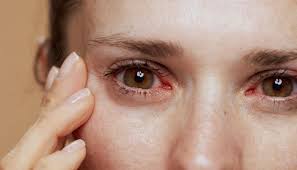
Blur that is more noticeable on waking and improves later in the day is a recognized Fuchs symptom. Glare, halos around lights and sensitivity to bright light can also interfere with vision, particularly in dim surroundings or at night. Some people have early findings without noticeable symptoms, while more advanced disease can cause persistent visual difficulty and pain. The common later-onset presentation often becomes symptomatic in middle or later adult life. These patterns are useful clues for an examination, but they do not establish the diagnosis at home. Describe any change to the clinician, who can connect symptoms with the examination findings. [3]
A useful symptom description separates clarity, comfort and the circumstances in which the problem occurs. For clarity, describe whether words look washed out, lights create troublesome glare, or one familiar task has become harder. For comfort, record whether there is soreness, grittiness or sensitivity to light. Then note the setting: after waking, during a work shift, outdoors, or while looking at an illuminated screen. Write ordinary examples rather than trying to translate everything into medical vocabulary. “I stopped reading the small labels on the kitchen shelf” gives the appointment a concrete starting point. Record questions as they arise so they do not depend on memory in the examination room. Keep the record brief enough to bring to the visit and discuss, and ask the clinician which changes should prompt earlier contact. [23]
Dry eye is one reason symptoms can be difficult to interpret. Problems with the tear film can produce burning, a scratchy sensation, redness, light sensitivity and blur. These overlap with complaints someone with corneal disease may report, even though tear-film dysfunction and endothelial dysfunction involve different processes. Assessment may include checking the quantity of tears, how quickly they evaporate and the eyelids. A diagnosis of Fuchs dystrophy therefore does not explain every uncomfortable eye automatically. Tell the eye professional about both discomfort and visual change so they can consider additional causes. If dry eye is also diagnosed, its treatment may improve comfort, but the purpose of that treatment should be made clear. Ask which problem each recommended product is intended to address and what response the clinician expects. [7]
A cataract can create a different source of blur, faded colors, halos and troublesome night vision. It is cloudiness of the lens inside the eye. Because the cornea and lens are separate parts of the focusing system, a person may need an assessment of both when vision changes. Cataract surgery removes the cloudy lens and replaces it with an artificial lens. That describes what the operation treats; it does not establish how much of a particular person’s blur comes from the lens. In the consultation, ask which findings support a cataract as the main cause of difficulty and whether other eye conditions could limit the expected result. Understanding the contribution of each condition is especially helpful before discussing any sequence of operations. [8]
When an eye change should not wait
Get prompt professional advice for new eye pain, redness or worsening vision, even without the emergency combinations described above. A new painful red eye deserves prompt assessment, including in someone who already has a longstanding eye diagnosis. Seek emergency care when redness comes with reduced or changed vision, pain on looking at light, a severe headache with nausea, unequal pupils or a significant injury. Contact lens wearers should obtain urgent assessment for a red eye because infection is one possible cause. Do not use a previous diagnosis to explain away a new pattern or wait for a routine follow-up appointment to ask about severe symptoms. The clinician assessing the new problem needs to know about Fuchs dystrophy, any eye operation and current eye medicines. That background helps assessment, but an established condition does not remove the need to investigate an acute change. [9]
Flashes of light, a sudden increase in floaters, or a dark curtain or shadow across vision are another reason to seek eye care immediately. These can occur with retinal detachment, a problem at the back of the eye that requires urgent treatment. Floaters are moving spots, threads or squiggly shapes in the visual field; a marked new shower of them matters even if the eye is not painful. A curtain can cover part of the view rather than causing complete loss of sight. Do not wait to see whether such symptoms follow the familiar morning pattern of corneal blur. Describe when they began and whether one or both eyes seem affected, but arranging assessment takes priority over keeping a detailed symptom diary. [10]
How assessment and monitoring fit together
A slit lamp is a microscope with a bright light that lets the clinician inspect the cornea. Additional tests answer different questions. Pachymetry measures corneal thickness, while specular microscopy provides a view of the endothelial layer. Visual acuity testing measures how clearly a person can read the test targets. These findings help the eye professional describe the cornea and the effect on sight. Ask which tests were performed and what each result contributes to the assessment. A cell image and a reading-chart result are different kinds of information, so it is helpful to hear how the clinician brings them together. Keep the date and the eye tested attached to any result you receive, especially if care will be shared between different clinics. Request a plain-language explanation of unfamiliar terms in the report. [4]
A complete eye examination may include reading an eye chart, testing pupil responses, measuring eye pressure and examining the inside of the eye after the pupils are dilated. Those parts of the visit help place a corneal finding in the context of overall eye health. Dilation gives the clinician a better view of internal structures; it does not replace examining the cornea. After dilating drops, vision may be blurred and light sensitivity may last for several hours. Arrange a safe way home and bring sunglasses if helpful. Ask what tests are planned when booking so that work and transport arrangements are realistic. The frequency of future examinations depends on the individual’s eye health and risk, so a general screening interval should not be substituted for the plan agreed with the treating professional. [6]
A thickness result or cell count needs clinical interpretation. Cell counts can be difficult to obtain when disease obscures the endothelial image. Ask whether the measurement was reliable and how it compares with the examination and earlier results; an isolated number is not a self-treatment instruction. [24]
Fuchs dystrophy needs an individual monitoring plan rather than a universal timetable. Regular ophthalmologist review can assess changes in the cornea and whether treatment is needed. The amount of corneal damage and its effect on the person help shape the discussion. A diagnosis at one visit does not supply a reliable date for a future operation. Ask what the clinician wants to compare at the next review and which changes should trigger contact sooner. When transferring care, provide earlier reports if available so the new team can understand what has already been observed. Monitoring can include periods of observation without active treatment; the important point is to understand the purpose of the plan and how to obtain reassessment when circumstances change. [25]
Genes, family history and uncertainty
Fuchs dystrophy can run in families, but its inheritance is not the same in every family. MedlinePlus Genetics describes an unknown inheritance pattern in many cases and an autosomal dominant pattern in some families. A rare early-onset form can involve changes in the COL8A2 gene. These distinctions matter because a simple statement that every child of an affected parent has a fixed chance of developing the disease is too broad. A person may also have Fuchs dystrophy without a known family history. Family information is useful background for care, not a prediction of exactly when symptoms will start or how severe they will become. Tell the clinician about relatives with confirmed corneal diagnoses or transplants, while being clear when the relative’s actual diagnosis is unknown. [5]
Genetic counseling can help when family questions are difficult to resolve. A genetics professional reviews medical and family history, explains patterns of inheritance and considers whether testing would answer a meaningful question. The discussion may include what a result can show, what it cannot establish and how it might affect decisions for relatives. Counseling is intended to support informed choices rather than direct people toward a particular personal decision. Before an appointment, gather confirmed diagnoses if relatives are comfortable sharing them; a family story about “an eye operation” may be too vague to interpret. Ask the eye specialist whether a referral is appropriate for the question at hand. Having a family history does not mean every family member needs the same tests or that testing will provide a complete explanation. [14]
Treatment goals and safe everyday care
Treatment is guided by symptoms and the effect on the cornea. Someone with early findings may not need active treatment. For some symptomatic people, a clinician may recommend a specially formulated salt-based eye medicine to draw fluid from the cornea and reduce swelling. More significant disease may lead to discussion of corneal transplantation. The purpose of each option should be explicit: easing symptoms, addressing a damaged layer or supporting vision during daily activities. Ask what improvement is realistic and how the team will decide whether the approach is helping. Do not turn a description of salt-based treatment into a homemade eye preparation; choosing and using an eye medicine requires the appropriate sterile product and individual instructions. [26]
Eye products need to be sterile because substances placed in the eye bypass some of the body’s defenses. A natural, herbal or homeopathic label does not establish that a product is suitable for the eye. Avoid homemade mixtures and ask a clinician or pharmacist about unfamiliar products or claims. Keep the dropper tip from touching the eye, fingers, clothing or other surfaces, and wash hands before handling eye medicines. If a product causes unexpected pain, discharge, discomfort or a vision change, contact a health professional promptly for advice. Keep the packaging available so they can identify the product. Safety warnings and recalls are product-specific; a reassuring brand name or an online testimonial is not a substitute for checking the exact bottle when a concern arises. [12]
The prescription or dispensing label should explain which eye a medicine is for and how it should be used. Ask for a demonstration if the bottle is difficult to squeeze, the print is hard to read or hand movement makes aiming difficult. Drop aids or a different container may help, and a pharmacist or clinic nurse can advise. Storage and expiry after opening vary between products, so use the directions for the actual medicine rather than a rule remembered from another bottle. Do not share eye drops. If more than one eye medicine is prescribed, ask the team to explain their order and timing in a written plan. Arrange refills before the supply runs out, particularly when travelling or when collecting prescriptions requires help from someone else. [13]
Daily habits can reduce other threats to the cornea even though they do not replace care for Fuchs dystrophy. Use appropriate protective eyewear for activities involving tools, flying particles, chemicals or certain sports. Ordinary glasses are not automatically a substitute for the protection required by a particular task. If contact lenses are used, follow the prescribed cleaning, disinfection and storage instructions. A new problem after lens wear warrants professional advice rather than improvising a different cleaning solution. Tell the eye professional about work or hobbies with dust, chemical splash or injury risk so that protection can fit the activity. This conversation is about avoiding an additional injury or infection while living with a corneal condition, rather than promising that a lifestyle change will reverse endothelial disease. [20]
Fuchs dystrophy and cataract surgery
Cataract surgery can place additional stress on endothelial cells, so known Fuchs dystrophy needs to be part of planning. Depending on the findings, a surgeon may consider cataract surgery alone, a corneal procedure first, or combined or staged treatment. The appropriate sequence requires individual assessment. Tell the cataract team about the corneal diagnosis before finalizing a plan and ask how they have evaluated the risk of persistent corneal clouding afterward. [2]
A planning conversation can be organized around the problem to solve and the next decision. Ask what is expected to improve after each proposed procedure, what might remain troublesome and what would happen if the first step did not achieve the hoped-for result. If a combined procedure is discussed, ask what the combined approach involves for appointments, recovery and later glasses assessment. If separate procedures are discussed, ask why that order is being proposed and how the next step will be decided. Give the team practical information about work, caring responsibilities, transport and support at home. These are questions for a shared discussion, not a formula that determines which operation is appropriate. Request a written summary that names the proposed procedure and the clinician responsible for answering questions before consent.
Understanding DMEK, DSAEK and other corneal transplants
Endothelial keratoplasty replaces diseased inner corneal tissue with donor tissue. DMEK stands for Descemet membrane endothelial keratoplasty. Its graft contains Descemet’s membrane and the endothelial cells. DSAEK means Descemet stripping automated endothelial keratoplasty; its graft also includes a thin layer of supporting corneal stroma. Both aim to address endothelial failure while leaving much of the person’s own cornea in place. The tissue difference explains the different names, but it does not tell a reader which procedure is appropriate for their eye. The surgeon considers the condition and clarity of the cornea and other features of the individual case. Ask why a particular approach is being offered, what alternatives are relevant and how the team expects it to address the person’s main visual or comfort problem. [15]
At an educational level, these operations involve removing the affected inner layer, introducing donor tissue through a small opening and positioning it against the back of the cornea. An air or gas bubble helps hold the graft in place while it attaches. Anesthesia is planned with the team; the approach varies with the patient and the procedure. Instructions after surgery may include a particular head or body position, checks before discharge and an early review. These details need to come from the operating unit because a general description cannot supply an individual recovery protocol. Before the operation, ask how the team will manage any difficulty lying flat or staying in the requested position. Confirm who will take the patient home and how the first follow-up visit will be reached. [16]
A discussion of success should identify the outcome being measured. Ask whether the surgeon is describing a clearer cornea, better vision with glasses, less discomfort, graft survival, or a lower chance of needing another procedure. Those are related goals but different questions. When percentages are offered, ask what group they describe, over what period and how closely that group resembles the situation being discussed. Also ask which uncertainties remain despite a careful examination. This makes consent more informative than a single headline figure. A patient can reasonably want both a plain-language explanation and time to consider the practical implications. Bring a trusted supporter if desired, request accessible written information and ask how to obtain clarification afterward. A treatment decision should rest on the individual consultation rather than another person’s online surgical story.
A full-thickness transplant is called penetrating keratoplasty, or PK. It replaces a central section containing the layers of the cornea, rather than just the innermost layer. The donor tissue is secured with fine stitches. This is a different operation from DMEK or DSAEK and may be considered when the clinical circumstances require it. Donor corneas are screened before use, although screening cannot remove every possible risk. Ask why the extent of tissue replacement is appropriate and how it affects follow-up and recovery. A general description of a corneal transplant can refer to several procedures, so confirm the full name of the operation being discussed before comparing information from different sources. [28]
Possible problems after endothelial transplantation include graft detachment, rejection, infection, raised eye pressure and graft failure. Detachment means the donor layer has not remained attached as intended; the team may need another air or gas procedure, sometimes called rebubbling, to help position it. Rejection is an immune response to donor tissue, while failure means the graft is not functioning adequately. They are not interchangeable terms. Some problems can be treated, and another transplant is sometimes considered, but no result is guaranteed. Ask which complications the team checks for during follow-up and how symptoms should be reported. A successful early result does not make later changes unimportant. New redness, pain, sensitivity to light or reduced vision after a graft needs prompt specialist assessment rather than waiting for the next scheduled review. [27]
Recovery is a continuing part of treatment
Vision usually takes time to settle after a corneal transplant, and glasses or contact lenses may still be needed. The recovery period varies by the type of transplant and the individual eye, so a general timeline is not a promise. Follow-up visits allow the team to assess progress and address problems. Prescribed eye medicines may continue long term to reduce the risk of the graft failing. Use them as directed and obtain advice before making a change. Return to driving only when the clinician agrees and the applicable vision requirements are met. Plan work and transport around the team’s advice rather than an assumed date. Discharge instructions should cover expected symptoms, changes needing urgent assessment and how to contact the service. [17]
Aftercare instructions can include protecting the eye, avoiding rubbing or pressure and temporarily changing activities such as swimming, contact sports or heavy work. The instructions depend on the operation and the treating service. Ask for a written plan covering the eye shield, washing, drops, activity restrictions and review appointments. Someone whose job involves dust, lifting or physical contact should explain those tasks rather than simply asking when “work” can restart. Support at home may be needed for transport, reading instructions or handling medicines. Keep the discharge paperwork where it can be found quickly, and give a helper the practical information they need with the patient’s agreement. Do not adopt another hospital’s positioning or medicine schedule from the internet: the plan supplied by the operating team should guide recovery. [18]
Living with visual difficulty while care continues
Support with daily tasks can be useful whenever vision is interfering with life. Adjustable lighting, reduced glare, larger text, magnification and phone or computer accessibility settings may make particular tasks easier. The best arrangement depends on the task and the person’s vision; simply making a light brighter is not always the most comfortable solution. If difficulty persists, ask about vision rehabilitation. A specialist can help with reading devices, accessible technology, home organization and strategies for remaining independent. Needing this support does not require a person to stop discussing treatment options with the eye team. Describe the activities that matter most, such as reading medication labels, cooking, using transport or continuing a hobby. A practical goal gives the rehabilitation discussion more direction than a general request to “see better.” [19]
How to interpret research and new-treatment claims
Research into the biology of Fuchs dystrophy includes efforts to understand its genetic contributors. In a 2017 NEI-funded study, investigators compared genetic information from people with and without the condition and identified associations involving several regions, including TCF4. Such studies can suggest biological pathways and future research questions. They do not, on their own, show that a genetic result predicts a particular person’s future vision or that a new treatment is ready for routine use. When reading a report, check its date and whether it describes laboratory work, an association study or treatment tested in people. Keep the original research question in view. A discovery about why a disease occurs can be valuable without immediately changing the care offered in an eye clinic. [21]
Descemet stripping only, or DSO, removes a selected central area of affected tissue without adding donor cells. Moorfields describes this approach as under investigation and not routine there. Its possible role depends on careful selection and the ability of remaining cells to cover the treated area. Availability and practice vary, so it should not be presented as a universal replacement for transplantation. Ask a corneal specialist what evidence and local safeguards apply to any proposed newer approach. [11]
For a clinical study, ask what is being tested, why it might help and how it differs from available care. Find out which tests and visits are required, who oversees eye care during the study and whether usual treatment can continue. Discuss known and uncertain short-term and long-term risks, possible benefits and costs. Participation can create additional demands on work, transport and support at home, so those details belong in the conversation. The research team should explain what will happen and answer questions before a decision is made. A study listing or recruitment notice is an invitation to learn about research, not evidence that the intervention will help a particular person. Take the information to the treating eye professional if it is unclear how the proposed study relates to the current care plan. [22]
A practical way to prepare for the next appointment
Choose the two or three questions that would make the biggest difference to understanding the next step. One might concern the diagnosis, another a treatment choice, and another a practical barrier to following the plan. Bring the names of current eye products, details of prior operations and any previous reports the clinic has requested. If a question remains unanswered, ask whether it can be addressed now or needs another appointment or professional. At the end, write down the agreed next step, the expected way of receiving results and the person to contact if the plan becomes difficult to follow. This creates a useful record without trying to turn the patient into their own specialist. Clear explanations and a workable plan are worthwhile goals at every stage, including periods when no operation is being considered.
Questions that make a care discussion clearer
| Topic | Question to bring | Useful detail to record |
|---|---|---|
| Current findings | What is established, and what remains uncertain? | The explanation in ordinary language |
| Monitoring | What is the next review intended to assess? | Appointment and earlier-contact instructions |
| Treatment | What is this option intended to improve? | Expected benefit, limitations and alternatives |
| An operation | Why is this procedure or sequence being proposed? | Individual risks and recovery arrangements |
| Daily life | Which tasks would benefit from additional support? | A practical plan for the activities that matter |
Frequently asked questions
Does everyone with Fuchs dystrophy need a transplant?
No. Treatment depends on symptoms and examination findings. Early disease may need no active treatment, while more significant visual problems can lead to a discussion of surgery. [11]
Is morning blur enough to diagnose Fuchs dystrophy?
No. Morning blur is a recognized symptom, but a professional eye examination is needed to establish its cause. [3]
Will every child of an affected parent develop Fuchs dystrophy?
No. Inheritance varies. Some families have an autosomal dominant pattern, while the inheritance pattern is unknown in many cases. [5]
What is the difference between DMEK and DSAEK?
Both replace inner corneal tissue. DMEK uses Descemet’s membrane and endothelial cells; DSAEK also includes a thin layer of supporting stroma. [15]
Do eye drops cure Fuchs dystrophy?
No. Swelling-relief drops may help symptoms, but they do not cure the underlying dystrophy. Ask the eye specialist what the prescribed product should achieve and how it will be reviewed. [26]
Which symptoms need urgent attention after a corneal transplant?
Contact the care team immediately for a red or painful eye, light sensitivity, new blurred vision, flashes, sudden new floaters or a curtain across vision. [17]
Sources
- National Eye Institute. How the Eyes Work.
- Moorfields Eye Hospital. Fuchs’ Endothelial Corneal Dystrophy.
- National Eye Institute. Corneal Dystrophies.
- MedlinePlus Medical Encyclopedia. Fuchs dystrophy.
- MedlinePlus Genetics. Fuchs endothelial dystrophy.
- National Eye Institute. Get a Dilated Eye Exam.
- National Eye Institute. Dry Eye.
- National Eye Institute. Cataracts.
- NHS. Red eye.
- National Eye Institute. Retinal Detachment.
- Moorfields Eye Hospital. Fuchs’ dystrophy diagnosis and treatment.
- U.S. Food and Drug Administration. What You Should Know about Eye Drops.
- Moorfields Eye Hospital. How to use your eye drops.
- MedlinePlus Genetics. What happens during a genetic consultation?.
- University Hospitals Sussex NHS Foundation Trust. Endothelial Corneal Graft Surgery.
- Royal Free London NHS Foundation Trust. Having a DMEK or DSAEK corneal transplant.
- NHS. Cornea transplant.
- Leeds Teaching Hospitals NHS Trust. Corneal Graft Post Operative Advice.
- National Eye Institute. Low Vision.
- National Eye Institute. Corneal Conditions.
- National Eye Institute. NIH-funded team identifies genetic underpinnings of Fuchs dystrophy.
- National Eye Institute. Clinical Trials: Questions to Ask.
- National Eye Institute. Talk to Your Doctor About Eye Health.
- American Academy of Ophthalmology EyeWiki. Fuchs’ Endothelial Dystrophy.
- American Academy of Ophthalmology. Fuchs’ Dystrophy patient brochure.
- Manchester Royal Eye Hospital. Fuchs’ Corneal Endothelial Dystrophy.
- Moorfields Eye Hospital. Endothelial keratoplasty.
- Cambridge University Hospitals NHS Foundation Trust. Corneal transplantation.