

Sarcoidosis symptoms can raise questions across several specialties. This guide focuses on recognizing organ-related patterns, understanding urgent changes and interpreting the tests used to investigate them. For broader background, see our sarcoidosis overview.
Symptoms that need urgent assessment
Call your local emergency number for sudden loss of vision, sudden one-sided weakness or trouble speaking, a first seizure, or ongoing palpitations with chest pain, breathlessness or fainting. Stroke-like symptoms need emergency help even if they stop. Do not drive yourself. A seizure lasting more than five minutes, repeated seizures without recovery, or difficulty breathing afterward also requires emergency help. A painful or very red eye, light sensitivity, or new blurred vision needs urgent eye assessment. Do not wait for a routine sarcoidosis appointment or assume these changes are a flare. If concerning palpitations and associated symptoms have stopped, seek urgent assessment. These symptoms can have other serious causes. 3
Why the symptom pattern varies
Sarcoidosis can affect different organs, so there is no single set of symptoms that everyone experiences. One person may have a persistent cough; another may notice a skin change or an eye problem. Some people have no symptoms and first learn of a possible problem through a chest image obtained for another reason. The location of an abnormality matters as much as the number of symptoms. A short symptom list cannot establish which organs are involved, and a long list of common complaints does not establish sarcoidosis. 1
Pulmonary sarcoidosis does not follow the same course in every person. Inflammation may settle, continue over time or return after an interval. Persistent inflammation can damage lung tissue, but a previous diagnosis does not establish whether a new respiratory symptom represents active inflammation, an established change or another illness. The present pattern needs to be compared with the earlier clinical picture rather than inferred from the name of the disease. 15
Cough, breathlessness and chest discomfort
A persistent dry cough, wheezing, chest discomfort and shortness of breath with activity are possible pulmonary symptoms. Inflammation and changes in lung tissue can make breathing less efficient; stiffness and reduced lung capacity may contribute to difficulty with exertion. The first noticeable change may be needing to pause during an activity that previously felt easy, rather than breathlessness while sitting still. These experiences also occur in other respiratory and cardiovascular conditions, so their presence does not identify sarcoidosis. 6
Investigating a cough involves more than naming it dry or productive. Its persistence, associated breathing difficulty and the chest findings affect the next steps. When bronchoscopy is appropriate, a clinician may obtain tissue or wash fluid through the airways. Examining that fluid can help characterize inflammation and investigate infection. It does not provide a unique sarcoidosis signature. Tissue findings, imaging and the clinical history must still fit together before a respiratory symptom is attributed to this disease. 6
A reassuring chest examination or normal lung-function measurement does not exclude sarcoidosis. Symptoms can be substantial even when those findings are relatively unremarkable. Conversely, the amount of cough does not reliably measure the extent of lung disease. The British Thoracic Society emphasizes that assessment should account for symptoms that seem out of proportion to measured pulmonary impairment, rather than dismissing them because one test looks satisfactory. 7
Breathlessness that is greater than expected from the lung findings can prompt consideration of cardiac involvement or pulmonary hypertension, meaning raised pressure in the lung circulation. These possibilities require targeted evaluation; they cannot be separated by the sensation of breathlessness alone. The question is not simply whether a breathing test falls inside its reference range. Clinicians also consider whether that result adequately explains the person’s limitation and whether another part of the heart and lung system needs investigation. 7
Fatigue and general symptoms
Sarcoidosis may begin gradually or with more noticeable symptoms over a short period. Tiredness, fever, unintentional weight loss and enlarged lymph nodes are possible general features. They describe a systemic illness pattern rather than a particular organ diagnosis. Enlarged nodes may be noticed externally, but other enlarged nodes are found on imaging and cannot be felt. The absence of a visible lump therefore does not settle whether lymph nodes are involved. 5
Sarcoidosis-related fatigue can interfere with concentration, work and ordinary activities even when other aspects of the illness appear controlled. It is not equivalent to laziness, and its severity cannot be read directly from a single blood result. A fatigue questionnaire may help describe the impact and track change, but it does not establish which mechanism is responsible. The experience deserves assessment even when it is not accompanied by a striking scan abnormality. 11
Several contributors may coexist. Assessment can consider ongoing inflammation, treatment effects, poor sleep or sleep apnea, anemia, thyroid disease, diabetes and mood-related difficulties. Identifying one contributor does not mean that the rest of the experience is imaginary. Equally, fatigue alone does not prove that granulomatous inflammation has returned or that a particular organ is newly affected. Distinguishing these possibilities matters because the appropriate response depends on the cause, and simply labeling the symptom a flare can leave another treatable problem unrecognized. 11
Skin changes, scars and tender nodules
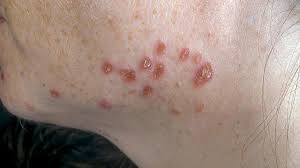
Skin sarcoidosis can produce bumps, thicker patches and changes in color or texture. Its appearance varies with skin tone, so an image showing a red lesion on one person is not a reliable template for everyone. Some affected areas are painless. A lesion therefore does not become unimportant simply because it does not itch or hurt, and color alone cannot determine what process is occurring beneath the surface. 10
Old scars, tattoos or piercing sites can become raised or otherwise change. A previously stable scar that develops a new texture is a clinical observation worth distinguishing from a new unrelated rash. It is not proof of sarcoidosis: examination may be needed to separate several possible causes. Skin findings can also lead clinicians to look beyond the skin, because visible disease and internal organ involvement are separate questions. The amount of visible skin change does not provide a dependable measure of disease elsewhere in the body. 10
Two named skin patterns illustrate why the type of lesion matters. Erythema nodosum produces tender nodules, typically on the shins, through inflammation in the fat beneath the skin. It is a nonspecific reaction that can accompany conditions other than sarcoidosis. It differs from lesions in which a biopsy demonstrates sarcoidal granulomas. Calling every sarcoidosis-associated bump a granuloma misses this distinction and may lead to unrealistic expectations about what a skin sample will show. 21
Lupus pernio describes persistent, often dusky thickened areas, particularly around the nose, cheeks or ears. Despite its name, it is not another name for systemic lupus. These patterns are clues for examination and selection of a suitable biopsy site, not diagnoses to make from a photograph. A clinician considers the shape, location and persistence of the lesion alongside other findings. The appearance of a skin lesion by itself cannot establish whether the lungs, heart or another internal organ is affected. 21
Ankles, joints, muscles and the Löfgren pattern
Joint involvement may cause pain, swelling and stiffness, often affecting joints in the lower limbs. The difficulty may be most obvious during walking or when moving after rest. Muscle involvement raises a different question: weakness or tender lumps may indicate a problem in muscle rather than inside the joint. Pain and weakness are not interchangeable descriptions, because an assessment of joint movement differs from an examination of muscle strength. 12
Bone abnormalities can be silent and discovered during investigation rather than announced by a distinctive pain. They also have alternative explanations. In a person receiving treatment, reduced bone strength associated with that treatment is a different issue from sarcoidosis directly involving bone. These distinctions help prevent all musculoskeletal complaints from being folded into one diagnosis. Examination and appropriately selected investigations establish whether the main problem concerns a joint, muscle, bone or another cause of limited movement; symptoms alone cannot reliably draw those boundaries. 12
Löfgren syndrome is an acute presentation associated with enlarged lymph nodes at both lung hila, tender shin nodules called erythema nodosum, and ankle arthritis or inflammation around the ankles. Fever may accompany the pattern. The chest component is an imaging finding, not something that can be established by feeling the ankles or inspecting the rash. An isolated swollen ankle or shin lump does not demonstrate the whole syndrome. Its recognition depends on how the findings fit together. 9
Sarcoidosis can also produce symptoms through nerves or hormone-regulating structures. Facial weakness, headaches or seizures represent different neurological patterns. Unusual thirst and increased urination can occur when hormone control of water balance is disturbed. These symptoms have other causes, and their presence alongside joint complaints does not prove a single explanation. In particular, sudden facial weakness must not be presumed to be a familiar nerve problem or a harmless manifestation of an existing sarcoidosis diagnosis. 9
Eye symptoms can occur without pain
Inflammation within the eye is called uveitis. It can cause blurred vision, floating spots, eye pain, redness or sensitivity to light. The combination varies according to the part of the eye involved. Inflammation farther back in the eye may not produce the obvious red, painful eye that people expect, and some ocular inflammation causes no noticeable symptoms. Absence of pain is therefore not a reliable way to exclude eye involvement. 25
Eye disease can become apparent before pulmonary symptoms do. This can complicate recognition when the person has no previous sarcoidosis diagnosis. An ophthalmic examination investigates the location and character of inflammation and its effect on sight, while clinicians consider other causes of granulomatous eye inflammation. Recognizing a compatible pattern is only part of that process. Visual symptoms should not be diagnosed from a symptom checklist, and apparently comfortable eyes do not establish that the structures inside them are unaffected. 25
Dryness, burning, redness and blurred vision are possible eye complaints in sarcoidosis, but they are not specific to it. A surface sensation such as dryness does not reveal whether inflammation is present deeper in the eye. Assessment connects the reported symptom with an examination rather than assuming that every uncomfortable eye has the same cause. New loss of vision needs emergency assessment, regardless of whether sarcoidosis has already been diagnosed. 27
Palpitations, faintness and heart function
Cardiac sarcoidosis can affect the heart’s electrical pathways, its muscle function or both. Electrical involvement may disturb the timing or transmission of the heartbeat; muscle involvement can impair the ability to pump effectively. These are related but different problems, which helps explain why one person’s main presentation may be a rhythm disturbance while another develops symptoms associated with impaired pumping. Neither presentation can be characterized fully by checking a pulse at home. 16
The heart may be affected even when disease elsewhere is not obvious, and cardiac involvement may cause few symptoms. This makes symptom-based reassurance unreliable. Specialist assessment can combine rhythm monitoring with imaging and the broader clinical picture. A test that examines electrical activity is answering a different question from one that examines heart structure or function. No single result is definitive in every case, so apparently conflicting information may require interpretation rather than choosing whichever finding seems most reassuring. 16
Clinicians consider cardiac sarcoidosis when otherwise unexplained findings include significant heart block, ventricular rhythm disturbances or reduced ventricular function. These are medical findings, not symptoms that a reader can identify accurately from a heartbeat sensation. The American Heart Association highlights the importance of integrating such clues with other evidence. A person does not need to fit a particular age profile before a concerning cardiac symptom deserves assessment. 24
Cardiac magnetic resonance imaging can show patterns of injury, but no late-enhancement pattern alone establishes sarcoidosis. Heart tissue sampling also has limitations because disease can be patchy: an unguided sample may miss an affected area. A negative biopsy therefore needs interpretation in context rather than being treated as an absolute exclusion. Diagnostic confidence may remain graded when tissue confirmation cannot be obtained. These uncertainties explain why specialists combine multiple kinds of information; they do not justify delaying emergency evaluation of collapse or serious ongoing cardiac symptoms. 24
Neurological symptoms and overlapping causes
Neurological involvement can produce numbness, weakness, stiffness of the limbs or problems with bladder control. The pattern depends on whether the brain, spinal cord or peripheral nerves are affected. A change in sensation is different from loss of strength, and difficulty walking can reflect more than one neurological mechanism. Examination aims to locate the problem within the nervous system before deciding which disease process is responsible. 23
Sarcoidosis can resemble other neurological conditions. A history of sarcoidosis elsewhere may make it one consideration, but it does not establish the cause of a new neurological deficit. Confirmation can be difficult because the most directly affected tissue may be a sensitive place to biopsy. This is one reason clinicians integrate examination, imaging and other evidence instead of relying on one symptom. New weakness or bladder-control difficulty should not be normalized as an expected flare, particularly when it is sudden, progressive or accompanied by other neurological changes. 23
Seizures, leg weakness and walking difficulties are among the possible neurological manifestations described by specialist centers. They represent changes in function rather than a characteristic feeling unique to sarcoidosis. A seizure, for instance, does not identify the underlying disease; it requires assessment of the event and its possible causes. Similarly, difficulty walking needs examination of strength, balance and other neurological functions, rather than being explained automatically by general tiredness. 26
Calcium changes and kidney problems
Sarcoidosis can alter vitamin D activation and increase calcium in the blood or urine. These are distinct measurements: increased urinary calcium and an elevated blood calcium result do not mean exactly the same thing. Excess calcium can contribute to kidney stones and kidney injury. A stone may draw attention to the issue, while impaired kidney function or a calcium abnormality may also be identified through testing without an obvious stone episode. 8
The clinical question extends beyond whether calcium is high. Clinicians consider the kidney findings, the degree and context of the abnormality, and other possible causes. A calcium result alone does not establish sarcoidosis, and normal blood calcium does not provide a complete assessment of every potential renal complication. Kidney disease may have more than one contributor. Medicines and supplements can also affect the interpretation and management of calcium balance, so this is an area for individualized assessment rather than self-directed changes based on a symptom article. 8
Less visible organ involvement
Liver involvement can produce abnormal blood tests without prominent symptoms; some people develop jaundice. The absence of abdominal discomfort therefore does not exclude liver involvement or establish normal function. An enlarged spleen and abnormalities in blood counts may also be part of the clinical picture. Blood-count changes need interpretation because they do not identify which mechanism is responsible simply by being present in someone with sarcoidosis. 14
Salivary-gland involvement may cause facial swelling or a dry mouth. Involvement of the nose and sinuses can cause persistent congestion, nosebleeds or reduced smell. These are familiar symptoms with many other possible causes, which is why persistence and accompanying findings matter. A blocked nose alone is not evidence of systemic sarcoidosis. When several less visible organ findings occur, examination and selected laboratory assessment help determine their significance. Their clinical importance cannot be estimated just by comparing how uncomfortable they feel with more obvious lung, skin or joint symptoms. 14
What the clinical history contributes
The clinical history helps define which explanations are plausible. Home and workplace exposures, medicines, previous illness and family history may change the interpretation of symptoms or imaging. Some medicines can be associated with sarcoid-like presentations, while exposure-related conditions can resemble the disease. These details guide investigation; they do not prove that a particular exposure caused sarcoidosis. A risk factor is not the same thing as a diagnostic finding. 13
Chest imaging and lung-function tests
Chest X-ray categories describe patterns in the chest, including enlarged lymph nodes, changes within lung tissue and fibrotic changes. They are often called radiographic stages, but the word stage can be misleading if understood as a compulsory sequence. People do not necessarily pass through each category in order. A chest classification is also not a score for the severity of disease throughout the body. 4
This distinction is important when a report appears reassuring while another organ is causing concern. Significant disease outside the lungs can occur regardless of the chest category. The chest image describes what is visible in that anatomical area; it does not examine the inside of the eyes, measure the heart’s electrical conduction or assess neurological function. Nor does a category alone explain all respiratory limitation. Clinical interpretation connects the radiographic pattern with symptoms and functional measurements, rather than using the stage number as a shortcut to a complete assessment. 4
Spirometry and gas-transfer testing examine different aspects of breathing. Spirometry measures airflow and the amount of air moved during breathing maneuvers; gas-transfer testing assesses how effectively gas passes from the lungs toward the blood. Imaging, by contrast, describes structure. These tests can complement one another because a structural finding and a functional measurement are not interchangeable descriptions of lung health. A clinician selects and interprets them according to the question being investigated. 19
Endobronchial ultrasound, often shortened to EBUS, uses an ultrasound-equipped bronchoscope to examine relevant lymph nodes within the chest and guide sampling. Its purpose differs from obtaining another ordinary chest image: it can help obtain tissue from a selected target. It is not a procedure that everyone with a cough needs. Whether further imaging, functional testing or sampling is useful depends on the existing findings and what additional information would clarify the diagnosis. A menu of available tests is not a universal testing schedule. 19
What biopsy can and cannot establish
When tissue sampling is needed, the choice of site matters. An accessible skin lesion may offer a suitable target and sometimes avoid sampling another organ. Bronchoscopy can obtain tissue from within the chest when that is appropriate. The aim is to obtain useful evidence with a procedure suited to the clinical question, not automatically to biopsy whichever organ has caused the greatest worry. Different suspected patterns may therefore lead to different sampling approaches. 17
Imaging can help define the structures involved, with CT providing more anatomical detail than a plain chest radiograph. In selected neurological investigations, lumbar puncture may supply information about inflammation and possible alternative causes. Neither the availability of a procedure nor a previously abnormal test means that everyone needs it. The value lies in what the result can add to the existing evidence. Tissue and other samples are interpreted alongside clinical findings, rather than being treated as self-explanatory proof detached from the rest of the assessment. 17
Granulomas are organized collections of inflammatory cells, and their presence does not by itself identify sarcoidosis. Even a non-necrotizing granuloma pattern must be interpreted with possible alternative causes in mind. Mycobacterial and fungal infections, exposure-related granulomatous disease and malignancy can enter the differential diagnosis. This is why a pathology report can be an important part of the evidence without being the entire answer. 20
Which alternatives need investigation depends on the individual pattern. Exposure history, imaging, the organ involved and other clinical findings influence the selection of tests. Excluding mimics does not mean ordering every possible infection or cancer test for every person. It means addressing realistic alternatives sufficiently to support the diagnosis being considered. A compatible symptom cluster and biopsy may still leave questions when another explanation fits. Keeping those questions open prevents an inflammatory label from prematurely ending the investigation and helps ensure that a different underlying disease is not overlooked. 20
Blood results and silent-organ assessment
Angiotensin-converting enzyme, usually abbreviated ACE, is sometimes measured during assessment, but it is not a stand-alone sarcoidosis test. Some people with sarcoidosis do not have an elevated result, and several other disorders can raise it. A normal ACE result cannot exclude the disease; a high result cannot independently confirm it. The result is meaningful only in relation to the clinical findings and the laboratory context. 22
Medicines and assay-related factors can affect interpretation. A number without that context should not be treated as a direct measure of how much sarcoidosis exists throughout the body. It also cannot locate an affected organ. A person with a concerning eye or cardiac symptom still needs the relevant assessment rather than reassurance based on ACE alone. Conversely, an isolated abnormal result needs explanation rather than automatic assignment to sarcoidosis. Medication decisions should remain with the treating clinician; readers should not stop prescribed medicines to influence a test result. 22
For people already diagnosed with sarcoidosis, the American Thoracic Society guideline addresses baseline assessment for organ involvement that may be silent. It suggests an eye examination, electrocardiogram and selected blood tests, including tests related to kidney function, liver involvement and blood counts. It recommends baseline serum calcium measurement. These recommendations concern evaluation after diagnosis, not a screening panel for every otherwise well person who has fatigue or a cough. 2
Several recommendations are conditional and rest on limited-certainty evidence. Routine echocardiography or ambulatory rhythm monitoring is not universally suggested for asymptomatic people with extracardiac sarcoidosis, although individual circumstances can justify their use. Investigating palpitations or fainting is a different situation from routine baseline screening in someone without cardiac symptoms. Baseline assessment is also distinct from deciding how often later testing should occur. New symptoms can change the clinical question even after an earlier assessment was satisfactory; follow-up should be guided by the actual disease pattern and findings. 2
When targeted investigations leave uncertainty
Neurosarcoidosis illustrates why diagnostic certainty can have levels. Consensus criteria distinguish possible, probable and definite diagnoses according to the combination of neurological findings, evidence supporting sarcoidosis and tissue confirmation where feasible and appropriate. These categories describe the strength of diagnostic evidence, not simply how severe the symptoms feel. They do not turn a symptom cluster into a diagnosis. 28
Neurological tests and imaging can show abnormalities without uniquely identifying their cause. Infection and malignancy remain among the alternatives that clinicians may need to consider. Biopsy is weighed in the context of feasibility and appropriateness; suspected neurological disease does not mean every person should undergo brain biopsy. Evidence from other affected tissue may contribute to the assessment. The resulting interpretation can remain qualified while still being clinically important. A report expressing uncertainty should be understood as a statement about the evidence available, rather than proof that the symptoms are minor or that further assessment has no value. 28
A targeted test is most useful when its purpose is clear. An electrocardiogram examines electrical activity, an echocardiogram assesses aspects of heart structure and function, and kidney or liver blood tests address different organ questions. A normal result in one domain does not answer a question in another. Likewise, an abnormal result may reveal that an organ needs attention without establishing why it is affected. 18
This article provides general education and cannot diagnose symptoms or replace individualized medical assessment. Original media are retained separately; their inclusion does not imply that every statement in them has been clinically reviewed for this revision.