

This guide brings together clinical explanations and practical questions for appointments, recovery, school, work and family life. Sources checked on 9 October 2026.
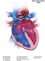
An atrial septal defect, or ASD, is an opening between the heart’s upper chambers that is present from birth. Blood commonly passes from the left atrium into the right atrium, sending extra blood through the right ventricle and lungs. A small opening may have little effect; a larger shunt can enlarge the right heart. Many children and some adults feel well, so a murmur or an incidental scan may be the first clue. Breathlessness during activity can develop. The important question is how the opening affects circulation, rather than whether symptoms are already obvious. [2]
ASD anatomy: why the exact diagnosis matters
The commonest type, a secundum ASD, lies in the central atrial septum near the oval depression called the fossa ovalis. A primum defect lies lower, beside the atrioventricular valves, and belongs to the atrioventricular septal defect, or AVSD, spectrum; abnormal valve anatomy may require treatment too. Sinus venosus defects occur near the venae cavae and frequently accompany pulmonary veins that drain abnormally into the right-sided circulation. An unroofed coronary sinus creates another, rarer atrial communication. These distinctions determine what must be imaged and repaired. A closure device designed for a central secundum opening cannot simply be applied to every communication between the atria. [3]
A patent foramen ovale, or PFO, is different. It is persistence of a flap-like fetal passage between overlapping septal tissues, rather than the tissue deficiency of a typical secundum ASD. PFOs are common and usually need no intervention. Decisions about closing a PFO after a carefully evaluated stroke follow a separate evidence base. Finding a PFO does not automatically explain neurological symptoms, and the adult PFO closure recommendations should not be extended to children. Keeping the terms separate prevents a common misunderstanding: an ASD assessment of right-heart volume loading is not interchangeable with a PFO-associated stroke evaluation. [4]
ASD also has more than one developmental explanation. Many cases are isolated, while others occur within a genetic syndrome or a family with congenital heart disease. The AHA genetics statement describes associations involving genes such as NKX2-5, GATA4 and TBX5; some inherited patterns also involve conduction abnormalities or limb differences. A family history therefore adds information beyond the size of the opening. Genetic evaluation is most useful when the clinical findings, associated abnormalities or family pattern suggest a specific question. It can inform relatives and reproductive counseling, but it does not provide a single genetic explanation for every ASD. [5]
How clinicians confirm the diagnosis and measure its effect
Transthoracic echocardiography, the ultrasound examination performed through the chest, is central to childhood assessment. A complete study examines the heart segment by segment, including venous connections, the atrial septum, valves and ventricles. Two-dimensional images show anatomy, while Doppler helps identify blood-flow direction. Measurements must be interpreted for a growing child rather than against adult dimensions. The 2024 ASE pediatric recommendations emphasize a comprehensive examination because identifying an opening is only part of the diagnosis. Associated abnormalities and the effect on chamber size can change both the interpretation and the next step. A focused image alone may leave those questions unanswered. [6]
In adults, the right ventricle deserves deliberate attention even when the left ventricle’s pumping measurement appears normal. The 2025 ASE right-heart guidance calls for assessment of chamber size, function and pressure-related findings together. One ultrasound number cannot describe the whole right heart. Enlargement, reduced contraction and evidence suggesting pulmonary hypertension answer different questions and should not be treated as synonyms. This distinction matters in ASD: extra blood volume can stretch a ventricle without proving severe pulmonary vascular disease. Consistent measurements over time help clinicians judge whether the circulation is stable or changing. [7]
Further imaging helps when the chest ultrasound cannot answer all the anatomical questions. Transesophageal echocardiography obtains views from the esophagus, close to the heart; cardiac magnetic resonance, or CMR, offers another way to examine the septum and surrounding structures. In a small comparative study of 20 adults being considered for device closure, CMR and transesophageal imaging agreed well on maximum defect size. That finding supports complementary options, rather than a rule that one test replaces the other in every patient. Image quality, the question being asked and the planned procedure determine which examination is useful. [8]
The notation Qp:Qs compares blood flow through the lungs with blood flow delivered to the body. A ratio of 1.5:1 means pulmonary flow is one-and-a-half times systemic flow. CMR can estimate this by measuring flow in major vessels; it need not rely only on tracing the small opening itself. A multicenter 4D-flow study found reproducible flow measurements, while direct measurement through the ASD was feasible in only 20 of 29 patients. This illustrates why specialists cross-check results. A visually impressive hole, one flow estimate and the amount of right-heart enlargement are related observations, but they are not interchangeable measurements. [9]
Observation and closure decisions in childhood
Some isolated secundum ASDs become smaller or close during childhood. In a longitudinal study of 200 children, smaller initial diameter and younger age at diagnosis predicted closure or regression. The study excluded additional congenital heart disease, syndromes and multiple or fenestrated defects, which limits how broadly its results can be applied. “Some close naturally” is therefore a reason for an appropriate observation plan, not a promise for a particular child. Follow-up compares the opening and its circulatory effects over time. A probability drawn from infants with isolated small defects should not be applied to a large defect diagnosed much later. [10]
Natural history can also run in the other direction. A separate serial-ultrasound study found enlargement in many of the 104 secundum defects it followed. Its device-size limits reflected technology available at that time, so those limits should not be reused as modern eligibility rules. The enduring lesson is that an ASD is not necessarily static. Growth of the child, change in defect dimensions and change in the surrounding tissue can affect future planning. This evidence helps explain why observation still involves reassessment, even when an initial consultation does not recommend immediate closure. [11]
The ACC’s 2026 pediatric guidance and its August 2026 updated ASD algorithm distinguish small, hemodynamically insignificant secundum defects from defects causing right-ventricular volume overload. Significant defects are generally closed during the preschool years; symptoms, associated conditions and anatomy can change timing. Selected small defects can be reviewed at intervals of several years, while stable patients after repair may also have widely spaced follow-up. These are decision-support pathways for defined patient groups, not a universal timetable. The document focuses on isolated lesions and explicitly leaves some complex anatomy and comorbidities outside its scope. It also acknowledges that pediatric follow-up practice contains evidence gaps. [12]
Adult closure: benefit depends on the circulation
An adult diagnosis does not mean that the opportunity for useful treatment has necessarily passed. An older randomized study in 473 adults aged over 40 found fewer events in a broad clinical composite that included recurrent pneumonia with surgical treatment than with medical observation. However, the trial selected patients with substantial shunts and excluded important hazards, including fixed pulmonary vascular resistance and left-ventricular dysfunction. Its message is about benefit in selected patients, not automatic closure at a particular birthday. Trial eligibility matters whenever results are translated into practice. Current decisions incorporate modern imaging, catheter techniques and the risks that made some patients unsuitable for that trial. [13]
The December 2025 ACC/AHA multisociety guideline replaces the 2018 adult guideline. For an unrepaired ASD with Qp:Qs at least 1.5 and right-ventricular enlargement, closure is recommended when pulmonary vascular resistance, or PVR, is at most 2 Wood units and significant left-ventricular disease is absent. Closure is reasonable with PVR above 2 but below 5. Selected patients starting at 5–8 may benefit if targeted pulmonary hypertension treatment lowers PVR below 5; benefit from fenestrated closure while PVR remains 5–8 is uncertain. All these pathways require specialist assessment of shunting, ventricular enlargement and left-heart disease. For evidence or strong suspicion of paradoxical embolism, where a clot crosses into arterial circulation, it recommends closure only in the absence of significant PAH or left-ventricular disease. It usually favors catheter closure over surgery for suitable isolated secundum defects. [14]
Another reason to pause before closure is a stiff or diseased left ventricle. The amount of atrial shunting depends partly on how readily each ventricle fills, as well as on the opening. An ASD can provide an outlet when left-atrial pressure is elevated; sealing it may raise filling pressure and worsen congestion. The ESC adult congenital guidance therefore calls for weighing complete closure, a deliberately fenestrated closure and leaving the defect open in relevant patients. A fenestration is a retained small passage, used for a specific physiological reason. The aim is a safer circulation, which is not always achieved by making the septum completely impermeable. [15]
Pulmonary hypertension and situations where closure may harm
Pulmonary hypertension means elevated pressure in the pulmonary circulation; pulmonary arterial hypertension is a specific category. The 2022 ESC/ERS definitions combine catheter-measured pressure, PVR and filling pressure to distinguish causes. In Eisenmenger physiology, severe pulmonary vascular disease reverses or makes the shunt bidirectional. Closing that communication is contraindicated. Closure is also contraindicated when severe PAH accompanies a small, incidental defect; this differs from a large, operable shunt. [16]
“Treat and repair” describes a specialist strategy in which pulmonary arterial hypertension is treated first, followed by reassessment of whether closure is appropriate. A multicenter observational report described favorable outcomes in carefully selected patients who responded to treatment and proceeded to closure; some other patients did not proceed because resistance failed to improve sufficiently. That selection is central to interpretation. A favorable result among responders cannot establish that every patient with ASD and PAH will become operable. The treatment response, remaining shunt, ventricular function and subsequent surveillance all matter, and medication may remain necessary after the anatomical repair. [17]
A September 2026 ERS update addresses PAH drug treatment and repeat right-heart catheterization during follow-up. Its focus includes a newer treatment pathway and reassessment when results would change management. It is not a blanket authorization to close an ASD. Advances in pulmonary vascular treatment and decisions about operability remain distinct questions. [16]
Temporary balloon occlusion can be used to explore the pressure response to closing an ASD, especially when left-heart filling is a concern. It is not a perfect rehearsal. A study of 42 adults found that wedge pressure during balloon occlusion could be higher than after device closure. That discrepancy cautions against treating one temporary measurement as an infallible prediction of pulmonary edema. The study was small and called for further work in higher-risk patients. Its practical contribution is to explain why specialists interpret a balloon test alongside the rest of the hemodynamic assessment, rather than as a simple pass-or-fail result. [18]
Catheter devices, surgery and evolving options
Device closure places an occluder across a suitable secundum opening through a catheter. Surgery closes the defect directly or with a patch and can address associated abnormalities. A multicenter comparison helped establish catheter treatment as an alternative, but it was not randomized: patients selected for the two approaches differed. That limitation remains relevant when comparing complication percentages across procedures. A patient requiring repair of abnormal pulmonary veins or valves is not equivalent to one with an isolated central opening and favorable rims. The choice starts with anatomy and the work required, rather than with a general claim that one procedure is best for everyone. [19]
High technical success does not mean zero risk. A prospective postapproval study followed 1,000 attempted Amplatzer device closures and documented rare serious events, including device embolization and cardiac erosion. The latter involves injury to adjacent cardiac tissue. The study’s selected population and two-year observation period are important limits when interpreting its findings. Preprocedure assessment therefore includes the supporting septal rims, neighboring structures and device fit. Follow-up also checks position and residual flow. A successful implantation is one milestone in care; it does not remove the need to investigate concerning symptoms or to complete the planned postprocedure assessments. [20]
The older shorthand that all sinus venosus defects require surgery now needs qualification. An international registry reported covered-stent correction as a catheter alternative in selected superior sinus venosus anatomy. This approach is different from placing a standard secundum occluder: it must also preserve the intended pulmonary venous pathway. Registry experience shows feasibility but cannot establish equivalence to surgery for every anatomy or over an entire lifetime. Detailed imaging, specialist experience and a plan for complications are essential. Inferior defects, unsuitable venous connections and other associated lesions still require their own assessment. [21]
Rhythm problems, follow-up and the long-term outlook
Atrial flutter and atrial fibrillation can occur before or after ASD repair. In a study of 213 adults undergoing surgery, pre-existing arrhythmia and older age helped identify patients with subsequent rhythm problems. Closing the opening does not necessarily reverse established electrical changes in the atria. This explains why rhythm history belongs in planning rather than being postponed until after repair. Palpitations may prompt electrocardiography or longer monitoring, depending on their pattern. A repair can address abnormal blood flow while a separate rhythm strategy addresses the electrical problem; these are related parts of care with different treatment targets. [22]
A Danish registry study also found that adults with ASD retained an increased risk of atrial fibrillation compared with matched population controls after closure. Stroke risk and pre-existing rhythm disease complicated the picture. Because this was observational research, the findings should not be read as proof that closure causes atrial fibrillation or that it lacks benefit. They show why anatomical repair and elimination of every future cardiovascular risk are different outcomes. Existing anticoagulation or rhythm treatment does not automatically become unnecessary when the defect is closed. Those decisions require assessment of the underlying indication and current clinical circumstances. [23]
An updated Dutch childhood-surgery cohort provides perspective over nearly half a century. The 2024 report found survival comparable with the general population and generally good exercise capacity and quality of life, while late supraventricular arrhythmias remained an important source of illness. This is encouraging without being a guarantee. It reflects people treated in a particular era and followed within one program, and it extends earlier reports from the same cohort rather than providing wholly independent evidence. Favorable long-term survival can coexist with a continuing need for assessment of rhythm, ventricular function and new symptoms as people age. [24]
Pregnancy and physical activity
The 2025 ESC pregnancy guideline describes uncomplicated repaired or unrepaired ASD without PAH as generally low risk, while recommending pre-pregnancy closure of large or hemodynamically significant defects. That category does not include every person carrying an ASD diagnosis. Pulmonary arterial hypertension, particularly Eisenmenger physiology, changes the risk substantially and warrants discussion with a Pregnancy Heart Team and a PH specialist. The guidance emphasizes informed, shared decisions and acknowledges very high risk in PAH. Pregnancy planning therefore considers the circulation, ventricular function, rhythm history and current treatment. The presence or absence of an old surgical scar cannot provide that assessment by itself. [25]
Physical activity advice should likewise match physiology. The 2025 AHA/ACC sports statement permits competitive participation after appropriate clinical evaluation for left-to-right shunts with minimal hemodynamic effect, while larger shunts, pulmonary hypertension, ventricular dysfunction or concerning symptoms require further assessment and shared decisions. Athletic training itself can enlarge heart chambers, so clinicians must distinguish training-related remodeling from shunt-related volume loading. Exercise capacity and rhythm findings can add information when the resting examination leaves uncertainty. Temporary restrictions after a procedure are another question from long-term eligibility. The ASD label alone cannot specify the right activity level for every child or adult. [26]
Everyday life, practical care, and family support
Making an ASD appointment useful
A useful appointment starts with the questions that matter most to the person living with ASD. Note any symptoms, when they began, whether they come and go, and what was happening when they occurred. Bring a current medicine and supplement list, relevant family history, and concerns about everyday life. Someone without symptoms can still ask what the next visit is intended to assess. [27]
Questions might include: What information is still missing? How could a test result change the plan? What are the benefits and drawbacks of the options being discussed? Which activities need individual advice? Request an explanation of unfamiliar terms and reliable reading material. Before leaving, identify the next step and write down any questions that remain unanswered. [27]
Keeping a clear medical summary
A short medical summary can help when a new clinician needs to understand years of care quickly. Boston Children’s Hospital recommends an accessible overview containing diagnoses, medicines, allergies, previous procedures and their dates, and specialist contacts. For someone with ASD, the procedure history should use the terminology recorded by the treating team rather than a remembered description such as a heart operation. [28]
Keep the summary linked to the fuller records it describes, and review it when care changes. Include communication preferences or accessibility needs that could affect an appointment. Patient portals and medical records departments can help fill gaps. This is general advice about organizing healthcare information; a personal summary supports communication but does not replace original reports or a clinician’s assessment. [28]
Planning for recovery at home
If a procedure is planned, recovery preparation should turn the hospital’s instructions into practical arrangements. For a child having catheter treatment, ask who will explain medicines, care of the access site, activity, and follow-up before discharge. Arrange for caregivers to hear the same instructions, and make sure they know which telephone number to use after the family returns home. [29]
Ask the team to distinguish expected changes from problems that need prompt contact, including concerns about bleeding, swelling, drainage, or fever. Confirm how and when recovery will be reviewed. Johns Hopkins’ guidance concerns children receiving catheter treatment; its timings should not be assumed to apply to surgery or to every adult. The treating service should provide the actual recovery plan, including any return-to-school advice. [29]
Making school support specific
School planning should describe a child’s actual needs. If appointments, recovery, symptoms, or prescribed care affect attendance or participation, discuss them with the school and healthcare team. A written plan can identify the responsible staff member, relevant medical instructions, emergency contacts, and arrangements for missed lessons. Include the child in the conversation in a way that fits their age and understanding. [30]
Review the plan when circumstances change, especially after a procedure or a change in clinical advice. Explain any specific participation guidance clearly so that staff can support inclusion. The American Academy of Pediatrics offers this approach for chronic conditions generally. An ASD diagnosis alone does not establish which educational services a child needs; those decisions require the child’s circumstances and the school’s assessment. [30]
Discussing work and caregiving demands
Work conversations become more useful when they describe the job itself. A desk role, lifting duties, long shifts, and professional driving raise different practical questions. Bring those details to the clinical team when discussing a return after treatment. Possible topics for an employer include appointment time, temporary duties, a gradual return, and a date to review whether the arrangement is working. [31]
Parents or other caregivers may need similar flexibility while supporting someone through hospital visits or recovery. The British Heart Foundation’s employment guidance applies broadly to heart conditions; it does not supply an ASD recovery deadline. Leave, benefits, disclosure requirements, and workplace protections differ by location and occupation. Seek appropriate local advice before making a major employment decision, particularly leaving a job. [31]
Asking about activity in practical terms
An activity discussion is more informative when it names the activity, its intensity, and the goal. Explain whether the question concerns playground games, a school team, recreational cycling, or competitive training. Describe any symptoms during exertion and any change in usual performance. The clinician can then consider the person’s heart findings and treatment history alongside the demands of that activity. [32]
If temporary limits are given after repair, ask which actions they cover and when they will be reassessed. Cleveland Clinic’s ASD information describes recovery restrictions, while its sports cardiology guidance emphasizes individual assessment. Neither provides a universal training plan for everyone with ASD. Written advice can help coaches and teachers understand the agreed plan without turning a diagnosis into an automatic ban on participation. [32]
Coordinating dental care
Tell the dentist about the ASD, any repair, the repair date, and information about an implanted device or patch. Regular oral care remains important. Antibiotics before dental treatment are recommended only for selected heart conditions and procedures; having ASD does not automatically mean that every dental visit needs preventive antibiotics. The cardiologist and dentist should confirm whether the individual meets the relevant criteria. Ask about endocarditis prevention for the planned dental procedure. [33]
Current guidance considers details such as recent device implantation and residual defects beside a patch or device. These require clinical interpretation. Ask for a written recommendation that the dental practice can retain, including whether it needs review later. Do not use another patient’s prescription or an old instruction as a substitute for that assessment. [33]
Helping children talk about worry
A child may worry about the next appointment, what a procedure will feel like, or being treated differently by friends. Start by asking what they understand and what is bothering them, then listen before offering reassurance. Predictable routines and time for enjoyable activities can help keep medical care from taking over every conversation. Give children choices where choices are genuinely available. [34]
Nemours’ advice on childhood stress is general guidance, rather than evidence that all children with ASD experience distress. The useful principle is to match support to the child. If worry is becoming persistent or difficult to manage, discuss it with their doctor and ask whether counseling would help. Feeling upset is a reason to offer support, not a failure to be brave. [34]
Including siblings and the wider family
When a child needs hospital care, brothers and sisters may need explanations of their own. They may misunderstand unfamiliar equipment, changes in family routines, or the attention being given to the patient. Use honest language suited to their age, invite questions, and make room for mixed feelings. Reassure them that an argument or something they said did not cause the hospitalization. [35]
Children’s Hospital of Philadelphia recommends protecting some individual time with siblings and helping them stay connected through calls, messages, drawings, or suitable visits. Friends and relatives may be able to help preserve familiar routines. These are general family-support approaches for hospitalization, not evidence that ASD always disrupts family life. Ask the hospital about additional sibling support when the family’s own resources feel stretched. [35]
Preparing for travel
Travel planning begins with the itinerary: destination, transport, altitude, planned activities, and access to medical care. Share relevant details with the congenital heart team, particularly when health has changed or treatment was recent. Carry a concise medical summary, contact details, and any device information the team recommends. If medicines are prescribed, discuss supplies for possible delays and keep essential medicines accessible during the journey. [36]
The Adult Congenital Heart Association also recommends considering insurance that addresses existing conditions and identifying appropriate care at the destination. Check any assistance or medical-equipment arrangements with the carrier before booking. These are planning principles for congenital heart disease generally. Whether a particular person with ASD needs additional precautions, oxygen assessment, or changes to travel plans requires advice from their own clinicians. [36]
Moving into adult congenital heart care
Transition is a gradual transfer of knowledge and responsibility as well as a change of clinic. Young people can start by learning the name of their heart condition, describing previous treatment, and asking some of their own questions. Over time, the plan can address arranging appointments, understanding prescribed medicines, and knowing how to contact the team when something is unclear. [37]
CDC guidance for congenital heart defects recommends starting this preparation during childhood and accounting for insurance and specialist access. Before pediatric care ends, establish which adult service will take over and how the transfer will happen. Someone who had an ASD repaired years earlier should bring that history into the discussion. The appropriate ongoing care depends on their clinical circumstances, rather than simply reaching a birthday. [37]
Planning pregnancy with specialist advice
Pregnancy planning is a reason to arrange an individualized review with a congenital heart specialist, ideally before pregnancy. If pregnancy has already begun, contact the care team promptly. An ASD label, a remembered childhood operation, or feeling well cannot by itself establish the care someone will need. The review should consider the person’s current heart condition and how pregnancy could affect it. [38]
The NHS recommends specialist assessment, review of medicines, and coordinated cardiac and maternity care for people with congenital heart disease. Bring the treatment history and medicine list, and ask who will organize follow-up and answer concerns between visits. Do not stop or change prescribed treatment without professional advice. Individual review should guide the next steps before pregnancy and throughout maternity care. [38]
Frequently asked questions
What is ASD?
ASD is an opening between the heart’s upper chambers, present from birth. [2]
Can an ASD close naturally?
Some small secundum defects shrink or close during childhood; follow-up checks their course. [10]
Does every ASD need closure?
No. Small, insignificant defects may be observed with an individualized follow-up plan. [12]
Are ASD and PFO the same?
No. A PFO is a persistent fetal flap passage; a typical secundum ASD lacks septal tissue. [4]
Does repair eliminate future rhythm problems?
No. Arrhythmias can persist or develop after repair. [22]
Are dental antibiotics always needed?
No. The dentist and cardiologist should confirm whether preventive antibiotics are indicated. [33]
Sources
- NHS: chest pain; NHS: breathlessness; NHS: stroke symptoms
- AHA: ASD overview
- ASE/SCAI: septal anatomy and imaging
- SCAI: PFO guidance
- AHA: congenital heart genetics
- ASE: pediatric echocardiography, 2024
- ASE: right-heart assessment, 2025
- Teo: CMR comparison study
- Chelu: 4D-flow MRI study
- Hanslik: childhood natural history
- McMahon: serial ASD growth study
- ACC: pediatric shunt guidance, 2026; ACC: updated ASD algorithm
- Attie: randomized adult closure trial
- ACC/AHA: adult congenital guideline, 2025
- ESC: adult congenital guidance, 2020
- ESC/ERS: pulmonary hypertension guidance, 2022; ERS: focused PAH treatment update, 2026
- Takaya: treat-and-repair study
- Nakagawa: balloon-occlusion study
- Du: device-versus-surgery comparison
- Turner: prospective device safety study
- Rosenthal: covered-stent registry
- Gatzoulis: adult rhythm outcomes
- Nyboe: Danish long-term registry
- Dutch childhood-repair cohort: fifty-year follow-up
- ESC: pregnancy guideline, 2025
- AHA/ACC: sports participation, 2025
- Mayo Clinic: ASD appointments
- Boston Children’s: medical summary
- Johns Hopkins: pediatric catheter recovery
- AAP: chronic conditions at school
- BHF: work and heart conditions
- Cleveland Clinic: ASD care; Cleveland Clinic: sports cardiology
- ADA: dental antibiotic prevention; AHA: cardiovascular device advisory, 2026
- Nemours: helping with childhood stress
- CHOP: support for siblings
- ACHA: travel planning
- CDC: transition to adult care
- NHS: congenital heart disease and pregnancy