Tetralogy of Fallot
Below is a comprehensive, structured report on Tetralogy of Fallot covering its definition, history, clinical features, causes, risk factors, complications, diagnosis, treatment options, prevention strategies, global statistics, recent research, and additional insights. This report is intended to be informative for both the general public and healthcare professionals.
1. Overview
What is Tetralogy of Fallot?
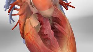
Tetralogy of Fallot (TOF) is a congenital heart defect characterized by four specific cardiac anomalies that impair the normal flow of blood through the heart. It is one of the most common cyanotic congenital heart diseases.
Detailed Definition
Tetralogy of Fallot comprises four anatomical abnormalities:
- Ventricular Septal Defect (VSD): A hole between the right and left ventricles.
- Pulmonary Stenosis: Narrowing of the pulmonary valve or right ventricular outflow tract, limiting blood flow to the lungs.
- Overriding Aorta: The aorta is positioned directly above the VSD, receiving blood from both ventricles.
- Right Ventricular Hypertrophy: Thickening of the right ventricular muscle due to increased workload.
Together, these defects cause deoxygenated blood to bypass the lungs and mix with oxygenated blood, leading to cyanosis (a bluish tint of the skin and mucous membranes).
Affected Body Parts/Organs
- Heart: The primary organ affected, with structural abnormalities involving the ventricles and outflow tracts.
- Circulatory System: Reduced pulmonary blood flow and mixing of oxygen-poor blood with oxygen-rich blood affect systemic oxygenation.
Prevalence and Significance
Tetralogy of Fallot is the most common cyanotic congenital heart defect, representing about 7–10% of all congenital heart diseases. Early surgical repair has greatly improved survival, but TOF remains a significant cause of pediatric morbidity and lifelong cardiac follow-up is required.
2. History & Discoveries
When and How Was Tetralogy of Fallot First Identified?
- Early Descriptions:
The condition was first described in the 1880s by Étienne-Louis Arthur Fallot, a French physician, who recognized the constellation of defects that now bears his name. - Modern Recognition:
Throughout the 20th century, advances in diagnostic imaging and cardiac surgery further elucidated the complex anatomy and improved management of TOF.
Who Discovered It?
- Étienne-Louis Arthur Fallot:
Fallot is credited with the first detailed description of the defect in 1888, establishing the foundation for understanding this congenital anomaly.
Major Discoveries and Breakthroughs
- Anatomical Clarification:
Detailed anatomical studies established the four hallmark features of TOF. - Surgical Repair:
The development of early surgical repair techniques in the 1950s, notably by Blalock, Taussig, and later by Lillehei and colleagues, revolutionized the prognosis. - Imaging Advances:
The advent of echocardiography, MRI, and CT scanning has enhanced preoperative planning and long-term follow-up.
Evolution of Medical Understanding
Medical understanding of TOF has evolved from initial descriptive pathology to sophisticated surgical and interventional strategies that provide near-normal life expectancy. Modern management now emphasizes early repair, long-term monitoring, and addressing residual lesions over the lifespan of the patient.
3. Symptoms
Early Symptoms vs. Advanced-Stage Symptoms
- Early Symptoms:
- Cyanosis (bluish skin, particularly of the lips and fingertips)
- “Tet spells” or hypercyanotic spells—sudden episodes of severe cyanosis and difficulty breathing, often triggered by crying or exertion in infants.
- Mild breathing difficulties and poor feeding in infants.
- Advanced-Stage Symptoms:
- Chronic fatigue and exercise intolerance.
- Recurrent respiratory infections.
- Clubbing of the fingers due to prolonged hypoxemia.
- In older children and adults, arrhythmias or heart failure symptoms may appear over time.
Common vs. Rare Symptoms
- Common Symptoms:
Cyanosis, tachypnea, and “tet spells” are typical in infants and young children. - Rare Symptoms:
Severe complications such as arrhythmias or right ventricular failure are less common in early childhood but may emerge later.
How Symptoms Progress Over Time
In TOF, symptoms often present early in life with cyanosis and feeding difficulties. With surgical repair, many children experience significant improvement, though they require lifelong monitoring for residual defects, arrhythmias, or pulmonary regurgitation that may develop with time.
4. Causes
Biological and Environmental Causes
- Biological Causes:
TOF is a congenital condition, meaning it occurs during fetal development. The precise cause is multifactorial, involving genetic mutations and disruptions in normal cardiac development. - Environmental Causes:
While no specific environmental factors cause TOF, maternal factors (such as diabetes or certain infections during pregnancy) have been implicated as potential contributors.
Genetic and Hereditary Factors
- Genetic Predisposition:
Genetic factors play a significant role; several genetic syndromes (e.g., DiGeorge syndrome) and mutations have been associated with TOF. A family history of congenital heart defects can also increase risk.
Known Triggers or Exposure Risks
- Developmental Triggers:
Abnormal signaling during heart development in the embryo, influenced by genetic and possibly maternal environmental factors, leads to the characteristic defects.
5. Risk Factors
Who Is Most at Risk?
- Age:
Since TOF is congenital, it is present at birth, with symptoms typically manifesting in infancy. - Genetic Factors:
Children born to parents with congenital heart disease or those with specific genetic syndromes have higher risk. - Maternal Health:
Maternal conditions such as diabetes may elevate the risk of congenital heart defects, including TOF.
Environmental, Occupational, and Genetic Factors
- Environmental:
There is no direct environmental cause postnatally, but maternal exposures during pregnancy may play a role. - Occupational:
Not applicable, as TOF is congenital. - Genetic:
Familial predisposition and associated genetic syndromes are significant risk factors.
Impact of Pre-existing Conditions
- Maternal Conditions:
Maternal diabetes and other metabolic disorders can impact fetal heart development. - Associated Syndromes:
Conditions like DiGeorge syndrome are associated with an increased incidence of TOF.
6. Complications
What Complications Can Arise from Tetralogy of Fallot?
- Pre-Surgical Complications:
Severe hypoxemia and “tet spells” may lead to developmental delays or organ dysfunction. - Post-Surgical Complications:
Residual defects, pulmonary regurgitation, arrhythmias, and right ventricular dysfunction. - Long-term Cardiac Issues:
Over time, some patients may develop heart failure, arrhythmias, or require re-interventions.
Long-term Impact on Organs and Overall Health
Without timely surgical intervention, chronic hypoxemia can impair growth and development, and damage other organs. Even after repair, lifelong follow-up is essential to manage and mitigate potential long-term cardiac complications.
Potential Disability or Fatality Rates
Before surgical repair was available, TOF carried a high mortality rate in infancy. Modern surgical techniques have greatly improved survival, though complications may still lead to disability and, in some cases, premature death if not managed appropriately.
7. Diagnosis & Testing
Common Diagnostic Procedures
- Prenatal Screening:
Fetal echocardiography can detect congenital heart defects including TOF. - Postnatal Clinical Examination:
Physical examination of cyanosis, heart murmurs, and signs of hypoxemia. - Echocardiography:
The primary diagnostic tool postnatally to visualize cardiac anatomy, assess the size of the VSD, degree of pulmonary stenosis, and right ventricular hypertrophy. - Electrocardiogram (ECG):
May show right ventricular hypertrophy and other electrical abnormalities. - Chest X-ray:
Can reveal a characteristic “boot-shaped” heart due to right ventricular hypertrophy.
Medical Tests
- Cardiac MRI:
Offers detailed imaging of heart structures and function, particularly useful in complex cases. - Cardiac Catheterization:
May be used for hemodynamic assessment and planning surgical interventions.
Early Detection Methods and Their Effectiveness
Early detection is critical; prenatal ultrasound and early postnatal echocardiography are highly effective in diagnosing TOF. Early intervention leads to improved outcomes and reduces the risk of complications.
8. Treatment Options
Standard Treatment Protocols
- Surgical Repair:
The definitive treatment is surgical correction, typically performed in infancy or early childhood. The most common procedure is complete repair, which includes closure of the VSD and relief of pulmonary stenosis (often via a transannular patch). - Palliative Procedures:
In some cases, temporary palliative shunts may be used if complete repair is not immediately possible. - Post-operative Care:
Includes medications to manage arrhythmias, diuretics for heart failure, and long-term follow-up with cardiology.
Medications, Surgeries, and Therapies
- Medications:
Pre- and post-operative management may include beta-blockers, diuretics, and antiarrhythmics. - Surgical Techniques:
Modern techniques include minimally invasive approaches and improved cardiopulmonary bypass methods. - Emerging Treatments:
Research continues on optimizing surgical outcomes, reducing re-interventions, and addressing residual lesions.
Emerging Treatments and Clinical Trials
- Advanced Imaging-Guided Surgery:
Use of intraoperative imaging and 3D modeling to enhance surgical precision. - Novel Surgical Approaches:
Investigational methods to reduce pulmonary regurgitation and improve right ventricular function. - Long-term Medical Management:
Clinical trials are ongoing to evaluate the best strategies for long-term management of repaired TOF, including pharmacologic interventions to prevent arrhythmias.
9. Prevention & Precautionary Measures
How Can Tetralogy of Fallot Be Prevented?
- Prevention of Congenital Heart Defects:
There is no known prevention for TOF as it is a congenital condition. However, maternal health optimization (e.g., controlling diabetes, avoiding teratogens) may reduce the risk of congenital defects in general. - Early Diagnosis and Intervention:
Prenatal care and early screening are essential to detect congenital heart defects early.
Lifestyle Changes and Environmental Precautions
- Maternal Health:
Encouraging proper nutrition, avoiding harmful substances during pregnancy, and regular prenatal check-ups. - Genetic Counseling:
For families with a history of congenital heart disease, counseling can provide insights into risks and potential preventive measures.
Vaccines or Preventive Screenings
- Preventive Screenings:
Routine prenatal ultrasounds and fetal echocardiography are key to early detection. There are no vaccines for TOF.
10. Global & Regional Statistics
Incidence and Prevalence Rates Globally
- Prevalence:
Tetralogy of Fallot is estimated to occur in approximately 3–5 per 10,000 live births, making it one of the most common cyanotic congenital heart diseases. - Regional Trends:
Incidence rates vary slightly by region, though it is observed worldwide. Developed countries typically have robust screening programs, while incidence in developing regions may be underreported.
Mortality and Survival Rates
- Mortality:
Before surgical advances, mortality was high; however, modern surgical repair has improved survival dramatically. Long-term survival rates exceed 90% in centers with advanced cardiac care. - Survival:
Lifelong follow-up is necessary, as some patients may experience complications later in life.
Country-wise Comparison and Trends
- Developed Countries:
High success rates with surgical repair and long-term management. - Developing Countries:
Outcomes may be poorer due to limited access to specialized cardiac care and delayed diagnosis.
11. Recent Research & Future Prospects
Latest Advancements in Treatment and Research
- Surgical Innovations:
Advances in minimally invasive and robotic-assisted surgery are being explored to reduce surgical morbidity. - Improved Long-term Management:
Research into better pharmacologic regimens to manage arrhythmias and ventricular dysfunction post-repair. - Genetic and Molecular Research:
Studies on the genetic basis of congenital heart disease may eventually lead to preventive strategies.
Ongoing Studies and Future Medical Possibilities
- Clinical Trials:
Ongoing trials are focused on refining surgical techniques, reducing residual defects, and improving long-term cardiac function. - Precision Medicine:
Integrating genetic data to individualize treatment and surveillance strategies. - Regenerative Therapies:
Early research into cardiac tissue engineering and stem cell therapy offers potential future avenues for repair and regeneration.
Potential Cures or Innovative Therapies Under Development
While there is no “cure” for congenital heart defects, innovative therapies—including improved surgical techniques and regenerative approaches—promise to enhance outcomes, reduce complications, and improve quality of life for individuals with TOF.
12. Interesting Facts & Lesser-Known Insights
Uncommon Knowledge About Tetralogy of Fallot
- Historical Milestone:
Named after Étienne-Louis Arthur Fallot, the syndrome was first systematically described in the 19th century and has since become a landmark in congenital cardiology. - Impact on Quality of Life:
Even with successful surgical repair, patients require lifelong monitoring due to the potential for late complications such as arrhythmias and right ventricular dysfunction. - Cultural Influence:
Advances in the treatment of TOF have dramatically changed the outlook for congenital heart disease, transforming a once-fatal condition into a manageable chronic disease.
Myths and Misconceptions vs. Medical Facts
- Myth: “Tetralogy of Fallot is always fatal in infancy.”
Fact: Modern surgical repair has improved survival rates dramatically, and many patients live into adulthood with good quality of life. - Myth: “Surgical repair is a one-time cure.”
Fact: While surgery corrects the defects, long-term follow-up is essential due to the risk of residual or progressive complications. - Myth: “There is no hope for preventing congenital heart disease.”
Fact: Although prevention is challenging, optimal maternal health and genetic counseling can reduce risk factors.
Impact on Specific Populations or Professions
- High-Risk Populations:
Newborns with congenital heart defects and families with a history of congenital cardiac anomalies. - Occupational Impact:
Pediatric cardiologists, cardiac surgeons, and specialized nurses play a critical role in the management and follow-up of TOF, reflecting the multidisciplinary nature of care. - Economic Impact:
Advances in TOF treatment have significantly reduced the long-term healthcare burden, though initial surgical and follow-up care remain resource-intensive.
References
- – Provides detailed information on the epidemiology, pathophysiology, and treatment of Tetralogy of Fallot.
- – Offers global statistics and public health guidelines on congenital heart diseases.
- – Summarizes recent clinical studies and advances in the management of Tetralogy of Fallot.
This report integrates current clinical knowledge and research findings to provide a detailed overview of Tetralogy of Fallot. Understanding its complex anatomy, clinical progression, and evolving treatment strategies is essential for optimizing patient care, guiding long-term management, and informing future research in congenital cardiology.