Dermatomyositis
Below is a comprehensive, structured report on Dermatomyositis covering its definition, history, clinical features, causes, risk factors, complications, diagnosis, treatment options, prevention strategies, global statistics, recent research, and additional insights. This report is designed to be informative for both the general public and healthcare professionals alike.
1. Overview
What is Dermatomyositis?
Dermatomyositis is a rare, chronic inflammatory disease characterized by distinctive skin rashes and progressive muscle weakness. It is classified as an idiopathic inflammatory myopathy with an autoimmune basis.
Detailed Definition
Dermatomyositis is an autoimmune condition that primarily affects the skin and skeletal muscles. It leads to muscle inflammation (myositis) and a characteristic violaceous (purple) rash, often on the eyelids, face, and upper body. Although the exact cause remains unknown, the disease is thought to result from an immune-mediated process triggered by genetic and environmental factors.
Affected Body Parts/Organs
- Muscles: Particularly proximal muscles (shoulders, hips) become weak and fatigued.
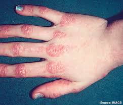
- Skin: A distinctive rash—often on the eyelids (heliotrope rash), knuckles (Gottron’s papules), and other sun-exposed areas—is a hallmark.
- Other Organs: In some cases, lung involvement (interstitial lung disease) or cardiac and gastrointestinal manifestations may occur.
Prevalence and Significance
Dermatomyositis is a rare disorder, with an estimated incidence of 0.5–0.6 per 100,000 people annually. Despite its rarity, it significantly impacts quality of life due to chronic muscle weakness, disability, and associated complications. Early diagnosis and treatment are critical to improving outcomes and reducing long-term morbidity.
2. History & Discoveries
When and How Was Dermatomyositis First Identified?
- Early Descriptions:
The condition was first described in the late 19th century. Early physicians recognized a link between skin changes and muscle weakness. - Modern Recognition:
Over the 20th century, improved clinical, histological, and immunological techniques helped define dermatomyositis as a distinct entity among inflammatory myopathies.
Who Discovered It?
- Historical Contributors:
Early pioneers in rheumatology and dermatology, including early case series published in the 1890s, contributed to recognizing the combination of myositis and characteristic skin findings. There is no single “discoverer,” but the collective work of clinicians and pathologists paved the way.
Major Discoveries and Breakthroughs
- Histopathology:
The identification of inflammatory infiltrates, perifascicular atrophy, and complement deposition in muscle biopsies clarified the diagnosis. - Immunological Insights:
Advances in autoantibody testing and understanding of the immune system’s role have refined classification and management. - Therapeutic Advances:
The introduction of corticosteroids and immunosuppressive agents in the mid-20th century significantly improved treatment outcomes.
Evolution of Medical Understanding
Medical understanding of dermatomyositis has evolved from descriptive clinical observations to a sophisticated appreciation of its autoimmune and inflammatory mechanisms. Today, research focuses on identifying specific autoantibodies and genetic factors that influence disease course and treatment response.
3. Symptoms
Early Symptoms vs. Advanced-Stage Symptoms
- Early Symptoms:
- Subtle, progressive muscle weakness (especially in the shoulders and hips)
- Mild fatigue and difficulty performing activities of daily living
- Initial skin changes, such as a faint reddish or purple discoloration around the eyes (heliotrope rash)
- Advanced-Stage Symptoms:
- Marked proximal muscle weakness leading to difficulty climbing stairs, rising from a chair, or lifting objects
- More pronounced and widespread skin rash, including Gottron’s papules over the knuckles and other extensor surfaces
- Systemic features such as weight loss, dysphagia, and, in some cases, interstitial lung disease
Common vs. Rare Symptoms
- Common Symptoms:
Proximal muscle weakness and characteristic skin rashes are typical. - Rare Symptoms:
Severe respiratory or cardiac involvement, significant dysphagia leading to nutritional deficits, or calcinosis (calcium deposits) are less frequently observed but indicate advanced disease.
How Symptoms Progress Over Time
Dermatomyositis often begins insidiously with subtle muscle weakness and skin changes. Without treatment, symptoms progress over weeks to months, resulting in significant functional impairment and systemic involvement. Early intervention can improve muscle strength and mitigate long-term complications.
4. Causes
Biological and Environmental Causes
- Biological Causes:
Although the exact etiology is unknown, dermatomyositis is considered an autoimmune disorder where the immune system mistakenly attacks muscle and skin tissues. - Environmental Causes:
Viral infections, ultraviolet light exposure, and certain medications have been proposed as environmental triggers that may initiate the autoimmune process in genetically predisposed individuals.
Genetic and Hereditary Factors
- Genetic Predisposition:
Genetic factors likely play a role, with certain HLA types and other genetic variations increasing susceptibility. Family history may contribute, though the disease is not directly inherited in a simple Mendelian pattern.
Known Triggers or Exposure Risks
- Infections:
Viral infections have been suggested as potential triggers. - Medications and Environmental Factors:
Certain drugs and environmental exposures (e.g., UV light) may precipitate disease onset in susceptible individuals.
5. Risk Factors
Who Is Most at Risk?
- Age:
Dermatomyositis can occur at any age but has peaks in childhood (juvenile dermatomyositis) and in adults, particularly between 40 and 60 years. - Gender:
There is a slight female predominance in adult-onset dermatomyositis. - Lifestyle:
Factors such as sun exposure may exacerbate skin symptoms. - Pre-existing Conditions:
Individuals with a history of autoimmune disorders or a family history of inflammatory diseases may have an increased risk.
Environmental, Occupational, and Genetic Factors
- Environmental:
Exposure to UV light is known to aggravate skin lesions. - Occupational:
No specific occupations have been conclusively linked, though jobs with high UV exposure could be contributory. - Genetic:
Certain genetic markers (e.g., specific HLA alleles) have been associated with an increased risk.
Impact of Pre-existing Conditions
Patients with pre-existing autoimmune conditions (such as rheumatoid arthritis or systemic lupus erythematosus) may be more susceptible to developing dermatomyositis or may experience more severe symptoms.
6. Complications
What Complications Can Arise from Dermatomyositis?
- Muscle Damage:
Chronic inflammation can lead to permanent muscle weakness and atrophy. - Calcinosis:
Deposition of calcium in soft tissues, particularly in juvenile dermatomyositis. - Interstitial Lung Disease:
Inflammation may extend to the lungs, leading to fibrosis and impaired respiratory function. - Cardiac Involvement:
Myocarditis and arrhythmias can occur in some patients. - Malignancy:
There is an association, particularly in adults, between dermatomyositis and certain cancers, necessitating malignancy screening.
Long-term Impact on Organs and Overall Health
Long-term complications can significantly impair mobility, respiratory and cardiac function, and overall quality of life. Chronic muscle weakness and pulmonary fibrosis, for example, can lead to substantial disability.
Potential Disability or Fatality Rates
While mortality directly due to dermatomyositis is relatively low with modern treatment, complications such as interstitial lung disease or associated malignancies can increase both morbidity and mortality rates.
7. Diagnosis & Testing
Common Diagnostic Procedures
- Clinical Evaluation:
Detailed history and physical examination, focusing on muscle strength, skin changes, and systemic symptoms. - Muscle Strength Testing:
Assessment of proximal muscle weakness through standardized scales. - Skin Examination:
Identification of characteristic rashes (heliotrope rash, Gottron’s papules).
Medical Tests
- Laboratory Tests:
- Creatine Kinase (CK): Elevated levels indicate muscle inflammation.
- Autoantibody Panels: Including anti-Jo-1 and other myositis-specific antibodies.
- Inflammatory Markers: ESR and CRP are typically elevated.
- Electromyography (EMG):
Evaluates muscle electrical activity to detect myopathic changes. - Muscle Biopsy:
Histopathological examination is the gold standard for confirming inflammation, muscle fiber necrosis, and perifascicular atrophy. - Imaging:
MRI can detect muscle inflammation and edema.
Early Detection Methods and Their Effectiveness
Early diagnosis relies on a combination of clinical evaluation, laboratory tests, and imaging studies. Timely detection is crucial for initiating treatment to prevent irreversible muscle damage and systemic complications.
8. Treatment Options
Standard Treatment Protocols
- Corticosteroids:
High-dose corticosteroids (e.g., prednisone) are typically the first-line treatment to reduce inflammation. - Immunosuppressive Agents:
Medications such as methotrexate, azathioprine, or mycophenolate mofetil are often used as steroid-sparing agents. - Biologic Therapies:
In refractory cases, biologics like rituximab or IVIG (intravenous immunoglobulin) may be employed. - Supportive Therapies:
Physical therapy and occupational therapy to maintain muscle strength and function. - Antimalarial Agents:
Sometimes used for their anti-inflammatory effects on the skin.
Medications, Surgeries, and Therapies
- Medications:
Corticosteroids remain the mainstay, complemented by immunosuppressants and, in some cases, IVIG. - Non-Pharmacologic Therapies:
Rehabilitation and physical therapy are critical for improving functional outcomes. - Emerging Treatments:
Newer biologic agents targeting specific inflammatory pathways (e.g., anti-TNF, anti-IL-6) are under investigation.
Emerging Treatments and Clinical Trials
- Biologic Agents:
Clinical trials are evaluating the efficacy of targeted biologics in reducing inflammation and preventing relapses. - Novel Immunomodulators:
Investigational drugs aimed at modulating the immune response and reducing muscle damage. - Personalized Medicine:
Research into genetic and molecular biomarkers may lead to more tailored treatment strategies in the future.
9. Prevention & Precautionary Measures
How Can Dermatomyositis Be Prevented?
- Primary Prevention:
There is no known method to prevent dermatomyositis, as its etiology is multifactorial and largely idiopathic. - Early Detection:
Regular medical evaluations for individuals with a family history of autoimmune diseases or persistent muscle weakness may facilitate early diagnosis. - Lifestyle Measures:
While specific lifestyle changes cannot prevent the disease, maintaining overall health through a balanced diet, regular exercise, and stress management may support immune regulation.
Lifestyle Changes and Environmental Precautions
- General Health:
Adopting a healthy lifestyle may help modulate immune responses, though evidence is limited regarding direct prevention of dermatomyositis. - Sun Protection:
For patients with photosensitive skin rashes, using sunscreen and protective clothing can reduce flare-ups.
Vaccines or Preventive Screenings
- Preventive Screenings:
Routine health check-ups and early evaluation of unexplained muscle weakness are recommended. - Vaccines:
There are no vaccines for dermatomyositis.
10. Global & Regional Statistics
Incidence and Prevalence Rates Globally
- Prevalence:
Dermatomyositis is a rare disease, with an estimated prevalence of 1–9 cases per 100,000 individuals. Juvenile dermatomyositis is even less common. - Regional Trends:
The disease is observed worldwide, though incidence may vary with geographic and ethnic factors.
Mortality and Survival Rates
- Mortality:
Mortality rates have improved with advances in treatment, though severe cases, particularly those with associated malignancy or interstitial lung disease, have higher mortality. - Survival:
With appropriate immunosuppressive therapy and supportive care, many patients achieve remission; however, relapses are common.
Country-wise Comparison and Trends
- Developed Countries:
Access to advanced diagnostic and treatment options has improved outcomes. - Developing Countries:
Limited resources may lead to delayed diagnosis and treatment, affecting overall prognosis.
11. Recent Research & Future Prospects
Latest Advancements in Treatment and Research
- Biologic Therapies:
Recent studies on biologic agents targeting specific cytokines (e.g., anti-TNF, anti-IL-6) show promise in refractory cases. - Immunomodulatory Research:
Advances in understanding the molecular and genetic mechanisms underlying dermatomyositis are guiding the development of targeted therapies. - Combination Therapies:
Investigations into optimizing combination regimens (e.g., corticosteroids plus immunosuppressants) aim to improve long-term outcomes.
Ongoing Studies and Future Medical Possibilities
- Clinical Trials:
Multiple clinical trials are ongoing to assess the safety and efficacy of new immunosuppressive and biologic agents. - Precision Medicine:
Research into biomarkers and genetic profiles may eventually allow for personalized treatment strategies. - Innovative Approaches:
New therapies, including mesenchymal stem cell treatments and novel immunomodulators, are under investigation.
Potential Cures or Innovative Therapies Under Development
While no cure exists for dermatomyositis, emerging therapies that more specifically target the autoimmune process may lead to longer remissions and improved quality of life. Ongoing research holds promise for more effective, individualized treatment regimens in the future.
12. Interesting Facts & Lesser-Known Insights
Uncommon Knowledge About Dermatomyositis
- Diagnostic Challenge:
Dermatomyositis is a challenging diagnosis due to its variable presentation and overlap with other autoimmune and neuromuscular disorders. - Association with Malignancy:
In adults, dermatomyositis may be a paraneoplastic syndrome, prompting thorough cancer screening. - Impact on Daily Life:
Chronic muscle weakness and skin changes can profoundly affect physical function and emotional well-being.
Myths and Misconceptions vs. Medical Facts
- Myth: “Dermatomyositis is just a skin condition.”
Fact: While the skin rash is characteristic, the disease also involves significant muscle inflammation and systemic involvement. - Myth: “It only affects children.”
Fact: Dermatomyositis occurs in both children (juvenile dermatomyositis) and adults, with different clinical courses. - Myth: “Once treated, the disease is cured.”
Fact: Dermatomyositis is often chronic and relapsing, requiring long-term management and follow-up.
Impact on Specific Populations or Professions
- High-Risk Populations:
Individuals with other autoimmune diseases or a family history of inflammatory conditions are at increased risk. - Occupational Impact:
Those in physically demanding professions may be significantly impacted by muscle weakness and fatigue. - Economic Impact:
The chronic nature of the disease, combined with the need for ongoing treatment and monitoring, poses a considerable economic burden on patients and healthcare systems.
References
- – Provides comprehensive information on the diagnosis, pathophysiology, and management of dermatomyositis.
- – Offers global data and public health guidelines on inflammatory myopathies.
- – Summarizes recent clinical studies and advancements in dermatomyositis research.
This report integrates current clinical knowledge and research findings to provide a detailed overview of dermatomyositis. Understanding its complex pathophysiology, varied clinical presentation, diagnostic strategies, treatment options, and future research directions is essential for optimizing patient care and improving long-term outcomes in this challenging autoimmune condition.